|
Axillary Nerve Dysfunction
Axillary nerve dysfunction is any disorder caused by damage to the axillary nerve. The axillary nerve is a branch of the brachial plexus that innervates the deltoid and teres minor muscles. This nerve can be injured or damaged in a variety of ways - penetrating injury such as knife or gunshot wounds, surgical trauma, stretch injury (common after motor cycle accidents), and various metabolic or rheumatic conditions that may cause focal disruption of the blood supply to the nerve. Axillary nerve dysfunction is often medically investigated with electromyography Electromyography (EMG) is a technique for evaluating and recording the electrical activity produced by skeletal muscles. EMG is performed using an instrument called an electromyograph to produce a record called an electromyogram. An electromyog ..., which can help localize the lesion to a particular portion of the nerve .. References External links {{DEFAULTSORT:Axillary Nerve Dysfunction Peripheral nervous system di ... [...More Info...] [...Related Items...] OR: [Wikipedia] [Google] [Baidu] |
Neurology
Neurology (from el, wikt:νεῦρον, νεῦρον (neûron), "string, nerve" and the suffix wikt:-logia, -logia, "study of") is the branch of specialty (medicine), medicine dealing with the diagnosis and treatment of all categories of conditions and disease involving the brain, the spinal cord and the peripheral nerves. Neurological practice relies heavily on the field of neuroscience, the scientific study of the nervous system. A neurologist is a physician specializing in neurology and trained to investigate, diagnose and treat neurological disorders. Neurologists treat a myriad of neurologic conditions, including stroke, seizures, movement disorders such as Parkinson's disease, autoimmune neurologic disorders such as multiple sclerosis, headache disorders like migraine and dementias such as Alzheimer's disease. Neurologists may also be involved in clinical research, clinical trials, and basic research, basic or translational research. While neurology is a nonsurgical sp ... [...More Info...] [...Related Items...] OR: [Wikipedia] [Google] [Baidu] |
Axillary Nerve
The axillary nerve or the circumflex nerve is a nerve of the human body, that originates from the brachial plexus (upper trunk, posterior division, posterior cord) at the level of the axilla (armpit) and carries nerve fibers from C5 and C6. The axillary nerve travels through the quadrangular space with the posterior circumflex humeral artery and vein to innervate the deltoid and teres minor. Structure The nerve lies at first behind the axillary artery, and in front of the subscapularis, and passes downward to the lower border of that muscle. It then winds from anterior to posterior around the neck of the humerus, in company with the posterior humeral circumflex artery, through the quadrangular space (bounded above by the teres minor, below by the teres major, medially by the long head of the triceps brachii, and laterally by the surgical neck of the humerus), and divides into an anterior, a posterior, and a collateral branch to the long head of the triceps brachii branch. * The ... [...More Info...] [...Related Items...] OR: [Wikipedia] [Google] [Baidu] |
Brachial Plexus
The brachial plexus is a network () of nerves formed by the anterior rami of the lower four cervical nerves and first thoracic nerve ( C5, C6, C7, C8, and T1). This plexus extends from the spinal cord, through the cervicoaxillary canal in the neck, over the first rib, and into the armpit, it supplies afferent and efferent nerve fibers the to chest, shoulder, arm, forearm, and hand. Structure The brachial plexus is divided into five ''roots'', three ''trunks'', six ''divisions'' (three anterior and three posterior), three ''cords'', and five ''branches''. There are five "terminal" branches and numerous other "pre-terminal" or "collateral" branches, such as the subscapular nerve, the thoracodorsal nerve, and the long thoracic nerve, that leave the plexus at various points along its length. A common structure used to identify part of the brachial plexus in cadaver dissections is the M or W shape made by the musculocutaneous nerve, lateral cord, median nerve, medial cord, and ... [...More Info...] [...Related Items...] OR: [Wikipedia] [Google] [Baidu] |
Deltoid Muscle
The deltoid muscle is the muscle forming the rounded contour of the human shoulder. It is also known as the 'common shoulder muscle', particularly in other animals such as the domestic cat. Anatomically, the deltoid muscle appears to be made up of three distinct sets of muscle fibers, namely the # anterior or clavicular part (pars clavicularis) # posterior or scapular part (pars scapularis) # intermediate or acromial part (pars acromialis) However, electromyography suggests that it consists of at least seven groups that can be independently coordinated by the nervous system. It was previously called the deltoideus (plural ''deltoidei'') and the name is still used by some anatomists. It is called so because it is in the shape of the Greek capital letter delta (Δ). Deltoid is also further shortened in slang as "delt". A study of 30 shoulders revealed an average mass of in humans, ranging from to . Structure Previous studies showed that the insertions of the tendons of the delto ... [...More Info...] [...Related Items...] OR: [Wikipedia] [Google] [Baidu] |
Teres Minor Muscle
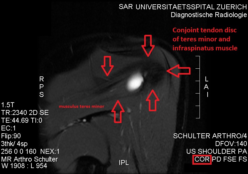
The teres minor (Latin ''teres'' meaning 'rounded') is a narrow, elongated muscle of the rotator cuff. The muscle originates from the lateral border and adjacent posterior surface of the corresponding right or left scapula and inserts at both the greater tubercle of the humerus and the posterior surface of the joint capsule. The primary function of the teres minor is to modulate the action of the deltoid, preventing the humeral head from sliding upward as the arm is abducted. It also functions to rotate the humerus laterally. The teres minor is innervated by the axillary nerve. Structure It arises from the dorsal surface of the axillary border of the scapula for the upper two-thirds of its extent, and from two aponeurotic laminae, one of which separates it from the infraspinatus muscle, the other from the teres major muscle. Its fibers run obliquely upwards and laterally; the upper ones end in a tendon which is inserted into the lowest of the three impressions on the greater tub ... [...More Info...] [...Related Items...] OR: [Wikipedia] [Google] [Baidu] |
Electromyography
Electromyography (EMG) is a technique for evaluating and recording the electrical activity produced by skeletal muscles. EMG is performed using an instrument called an electromyograph to produce a record called an electromyogram. An electromyograph detects the electric potential generated by muscle cells when these cells are electrically or neurologically activated. The signals can be analyzed to detect abnormalities, activation level, or recruitment order, or to analyze the biomechanics of human or animal movement. Needle EMG is an electrodiagnostic medicine technique commonly used by neurologists. Surface EMG is a non-medical procedure used to assess muscle activation by several professionals, including physiotherapists, kinesiologists and biomedical engineers. In Computer Science, EMG is also used as middleware in gesture recognition towards allowing the input of physical action to a computer as a form of human-computer interaction. Clinical uses EMG testing has a variety of ... [...More Info...] [...Related Items...] OR: [Wikipedia] [Google] [Baidu] |


