|
Atypical Polypoid Adenomyoma
Atypical polypoid adenomyoma (APA) is a rare benign tumour of the uterus. __TOC__ Pathology APAs are characterized by glands with abnormal shapes that: (1) often have squamous metaplasia, and (2) are surrounded by benign smooth muscle. Nuclear atypia, if present, is mild. The microscopic differential diagnosis includes endometrial carcinoma and endocervical adenocarcinoma. Image: Atypical polypoid adenomyoma - intermed mag.jpg , Intermed. mag. Image: Atypical polypoid adenomyoma - very high mag.jpg , Very high mag. See also *Adenomyoma *Cervical cancer Cervical cancer is a cancer arising from the cervix. It is due to the abnormal growth of cells that have the ability to invade or spread to other parts of the body. Early on, typically no symptoms are seen. Later symptoms may include abnormal ... References Benign neoplasms Noninflammatory disorders of female genital tract {{oncology-stub ... [...More Info...] [...Related Items...] OR: [Wikipedia] [Google] [Baidu] |
Micrograph
A micrograph or photomicrograph is a photograph or digital image taken through a microscope or similar device to show a magnified image of an object. This is opposed to a macrograph or photomacrograph, an image which is also taken on a microscope but is only slightly magnified, usually less than 10 times. Micrography is the practice or art of using microscopes to make photographs. A micrograph contains extensive details of microstructure. A wealth of information can be obtained from a simple micrograph like behavior of the material under different conditions, the phases found in the system, failure analysis, grain size estimation, elemental analysis and so on. Micrographs are widely used in all fields of microscopy. Types Photomicrograph A light micrograph or photomicrograph is a micrograph prepared using an optical microscope, a process referred to as ''photomicroscopy''. At a basic level, photomicroscopy may be performed simply by connecting a camera to a microscope, th ... [...More Info...] [...Related Items...] OR: [Wikipedia] [Google] [Baidu] |
H&E Stain
Hematoxylin and eosin stain ( or haematoxylin and eosin stain or hematoxylin-eosin stain; often abbreviated as H&E stain or HE stain) is one of the principal tissue stains used in histology. It is the most widely used stain in medical diagnosis and is often the gold standard. For example, when a pathologist looks at a biopsy of a suspected cancer, the histological section is likely to be stained with H&E. H&E is the combination of two histological stains: hematoxylin and eosin. The hematoxylin stains cell nuclei a purplish blue, and eosin stains the extracellular matrix and cytoplasm pink, with other structures taking on different shades, hues, and combinations of these colors. Hence a pathologist can easily differentiate between the nuclear and cytoplasmic parts of a cell, and additionally, the overall patterns of coloration from the stain show the general layout and distribution of cells and provides a general overview of a tissue sample's structure. Thus, pattern recogniti ... [...More Info...] [...Related Items...] OR: [Wikipedia] [Google] [Baidu] |
Pathology
Pathology is the study of the causes and effects of disease or injury. The word ''pathology'' also refers to the study of disease in general, incorporating a wide range of biology research fields and medical practices. However, when used in the context of modern medical treatment, the term is often used in a narrower fashion to refer to processes and tests that fall within the contemporary medical field of "general pathology", an area which includes a number of distinct but inter-related medical specialties that diagnose disease, mostly through analysis of tissue, cell, and body fluid samples. Idiomatically, "a pathology" may also refer to the predicted or actual progression of particular diseases (as in the statement "the many different forms of cancer have diverse pathologies", in which case a more proper choice of word would be " pathophysiologies"), and the affix ''pathy'' is sometimes used to indicate a state of disease in cases of both physical ailment (as in cardiomy ... [...More Info...] [...Related Items...] OR: [Wikipedia] [Google] [Baidu] |
Tumour
A neoplasm () is a type of abnormal and excessive growth of tissue. The process that occurs to form or produce a neoplasm is called neoplasia. The growth of a neoplasm is uncoordinated with that of the normal surrounding tissue, and persists in growing abnormally, even if the original trigger is removed. This abnormal growth usually forms a mass, when it may be called a tumor. ICD-10 classifies neoplasms into four main groups: benign neoplasms, in situ neoplasms, malignant neoplasms, and neoplasms of uncertain or unknown behavior. Malignant neoplasms are also simply known as cancers and are the focus of oncology. Prior to the abnormal growth of tissue, as neoplasia, cells often undergo an abnormal pattern of growth, such as metaplasia or dysplasia. However, metaplasia or dysplasia does not always progress to neoplasia and can occur in other conditions as well. The word is from Ancient Greek 'new' and 'formation, creation'. Types A neoplasm can be benign, potentially ma ... [...More Info...] [...Related Items...] OR: [Wikipedia] [Google] [Baidu] |
Uterus
The uterus (from Latin ''uterus'', plural ''uteri'') or womb () is the organ in the reproductive system of most female mammals, including humans that accommodates the embryonic and fetal development of one or more embryos until birth. The uterus is a hormone-responsive sex organ that contains glands in its lining that secrete uterine milk for embryonic nourishment. In the human, the lower end of the uterus, is a narrow part known as the isthmus that connects to the cervix, leading to the vagina. The upper end, the body of the uterus, is connected to the fallopian tubes, at the uterine horns, and the rounded part above the openings to the fallopian tubes is the fundus. The connection of the uterine cavity with a fallopian tube is called the uterotubal junction. The fertilized egg is carried to the uterus along the fallopian tube. It will have divided on its journey to form a blastocyst that will implant itself into the lining of the uterus – the endometrium, where it will ... [...More Info...] [...Related Items...] OR: [Wikipedia] [Google] [Baidu] |
Gland
In animals, a gland is a group of cells in an animal's body that synthesizes substances (such as hormones) for release into the bloodstream (endocrine gland) or into cavities inside the body or its outer surface (exocrine gland). Structure Development Every gland is formed by an ingrowth from an epithelial surface. This ingrowth may in the beginning possess a tubular structure, but in other instances glands may start as a solid column of cells which subsequently becomes tubulated. As growth proceeds, the column of cells may split or give off offshoots, in which case a compound gland is formed. In many glands, the number of branches is limited, in others (salivary, pancreas) a very large structure is finally formed by repeated growth and sub-division. As a rule, the branches do not unite with one another, but in one instance, the liver, this does occur when a reticulated compound gland is produced. In compound glands the more typical or secretory epithelium is found forming t ... [...More Info...] [...Related Items...] OR: [Wikipedia] [Google] [Baidu] |
Squamous Metaplasia
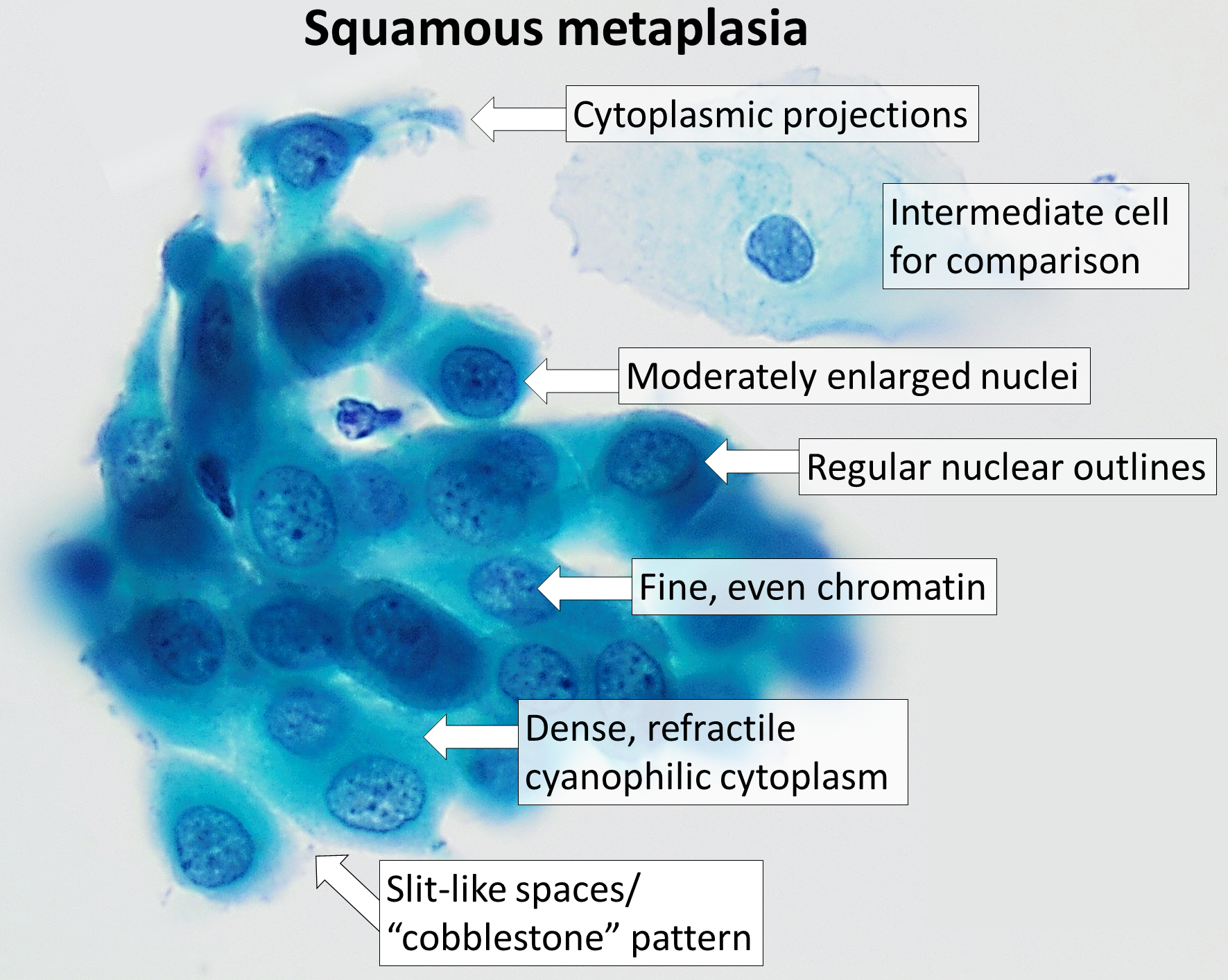
Squamous metaplasia is a benign non-cancerous change (metaplasia) of surfacing lining cells (epithelium) to a squamous morphology. Location Common sites for squamous metaplasia include the bladder and cervix. Smokers often exhibit squamous metaplasia in the linings of their airways. These changes don't signify a specific disease, but rather usually represent the body's response to stress or irritation. Vitamin A deficiency or overdose can also lead to squamous metaplasia. Uterine cervix In regard to the cervix, squamous metaplasia can sometimes be found in the endocervix, as it is composed of simple columnar epithelium, whereas the ectocervix is composed of stratified squamous non-keratinized epithelium.Kumar, Vinay; Abbas, Abul K.; Fausto, Nelson; & Mitchell, Richard N. (2007) ''Robbins Basic Pathology'' (8th ed.). Saunders Elsevier. pp. 716-720 Significance Squamous metaplasia may be seen in the context of benign lesions (e.g., atypical polypoid adenomyoma), chronic irri ... [...More Info...] [...Related Items...] OR: [Wikipedia] [Google] [Baidu] |
Smooth Muscle
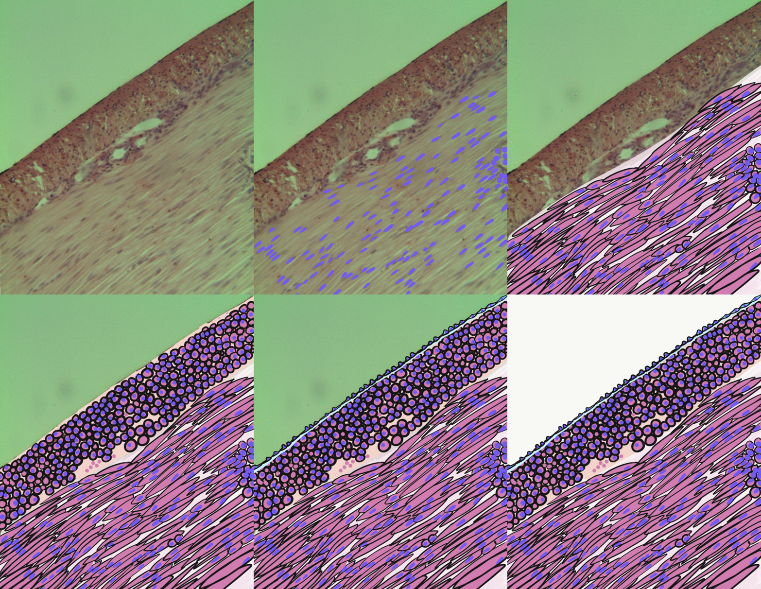
Smooth muscle is an involuntary non-striated muscle, so-called because it has no sarcomeres and therefore no striations (''bands'' or ''stripes''). It is divided into two subgroups, single-unit and multiunit smooth muscle. Within single-unit muscle, the whole bundle or sheet of smooth muscle cells contracts as a syncytium. Smooth muscle is found in the walls of hollow organs, including the stomach, intestines, bladder and uterus; in the walls of passageways, such as blood, and lymph vessels, and in the tracts of the respiratory, urinary, and reproductive systems. In the eyes, the ciliary muscles, a type of smooth muscle, dilate and contract the iris and alter the shape of the lens. In the skin, smooth muscle cells such as those of the arrector pili cause hair to stand erect in response to cold temperature or fear. Structure Gross anatomy Smooth muscle is grouped into two types: single-unit smooth muscle, also known as visceral smooth muscle, and multiunit smooth muscle. ... [...More Info...] [...Related Items...] OR: [Wikipedia] [Google] [Baidu] |
Nuclear Atypia
Nuclear atypia refers to abnormal appearance of cell nuclei. It is a term used in cytopathology and histopathology. Atypical nuclei are often pleomorphic. Nuclear atypia can be seen in reactive changes, pre-neoplastic changes and malignancy. Severe nuclear atypia is, in most cases, considered an indicator of malignancy. See also * Arias-Stella reaction *NC ratio The nuclear-cytoplasmic ratio (also variously known as the nucleus:cytoplasm ratio, nucleus-cytoplasm ratio, N:C ratio, or N/C) is a measurement used in cell biology. It is a ratio of the size (i.e., volume) of the cell nucleus, nucleus of a cell ... * Nuclear pleomorphism {{pathology-stub Pathology ... [...More Info...] [...Related Items...] OR: [Wikipedia] [Google] [Baidu] |
Differential Diagnosis
In healthcare, a differential diagnosis (abbreviated DDx) is a method of analysis of a patient's history and physical examination to arrive at the correct diagnosis. It involves distinguishing a particular disease or condition from others that present with similar clinical features. Differential diagnostic procedures are used by clinicians to diagnose the specific disease in a patient, or, at least, to consider any imminently life-threatening conditions. Often, each individual option of a possible disease is called a differential diagnosis (e.g., acute bronchitis could be a differential diagnosis in the evaluation of a cough, even if the final diagnosis is common cold). More generally, a differential diagnostic procedure is a systematic diagnostic method used to identify the presence of a disease entity where multiple alternatives are possible. This method may employ algorithms, akin to the process of elimination, or at least a process of obtaining information that shrinks the "p ... [...More Info...] [...Related Items...] OR: [Wikipedia] [Google] [Baidu] |
Endometrial Carcinoma
Endometrial cancer is a cancer that arises from the endometrium (the lining of the uterus or womb). It is the result of the abnormal growth of cells that have the ability to invade or spread to other parts of the body. The first sign is most often vaginal bleeding not associated with a menstrual period. Other symptoms include pain with urination, pain during sexual intercourse, or pelvic pain. Endometrial cancer occurs most commonly after menopause. Approximately 40% of cases are related to obesity. Endometrial cancer is also associated with excessive estrogen exposure, high blood pressure and diabetes. Whereas taking estrogen alone increases the risk of endometrial cancer, taking both estrogen and a progestogen in combination, as in most birth control pills, decreases the risk. Between two and five percent of cases are related to genes inherited from the parents. Endometrial cancer is sometimes loosely referred to as "uterine cancer", although it is distinct from other form ... [...More Info...] [...Related Items...] OR: [Wikipedia] [Google] [Baidu] |
Adenomyoma
Adenomyoma is a tumor (''-oma'') including components derived from glands (''adeno-'') and muscle (''-my-''). It is a type of complex and mixed tumor. Uterine adenomyoma In obstetrics and gynecology contexts, it is a form of adenomyosis that forms a mass or growth around the tissue of the inner uterus. Most cases of adenomyosis are non-symptomatic. However, it may present with dysmenorrhea and pelvic pain. In the case of juvenile cystic adenomyoma, laparoscopic enucleation results in a statistically and clinically significant reduction in dysmenorrhea, ease in any chronic pelvic pain, and low risk of recurrence. See also *Atypical polypoid adenomyoma *Adenomyosis Adenomyosis is a medical condition characterized by the growth of cells that proliferate on the inside of the uterus (endometrium) atypically located among the cells of the uterine wall (myometrium), as a result, thickening of the uterus occurs. A ... References External links Gynaecological neoplasia {{ ... [...More Info...] [...Related Items...] OR: [Wikipedia] [Google] [Baidu] |


.jpg)


