|
Arthroscopy
Arthroscopy (also called arthroscopic or keyhole surgery) is a minimally invasive surgical procedure on a joint in which an examination and sometimes treatment of damage is performed using an arthroscope, an endoscope that is inserted into the joint through a small incision. Arthroscopic procedures can be performed during ACL reconstruction. The advantage over traditional open surgery is that the joint does not have to be opened up fully. For knee arthroscopy only two small incisions are made, one for the arthroscope and one for the surgical instruments to be used in the knee cavity. This reduces recovery time and may increase the rate of success due to less trauma to the connective tissue. It has gained popularity due to evidence of faster recovery times with less scarring, because of the smaller incisions. Irrigation fluid (most commonly 'normal' saline) is used to distend the joint and make a surgical space. The surgical instruments are smaller than traditional instruments. ... [...More Info...] [...Related Items...] OR: [Wikipedia] [Google] [Baidu] |
Arthroscope
Arthroscopy (also called arthroscopic or keyhole surgery) is a minimally invasive surgical procedure on a joint in which an examination and sometimes treatment of damage is performed using an arthroscope, an endoscope that is inserted into the joint through a small incision. Arthroscopic procedures can be performed during ACL reconstruction. The advantage over traditional open surgery is that the joint does not have to be opened up fully. For knee arthroscopy only two small incisions are made, one for the arthroscope and one for the surgical instruments to be used in the knee cavity. This reduces recovery time and may increase the rate of success due to less trauma to the connective tissue. It has gained popularity due to evidence of faster recovery times with less scarring, because of the smaller incisions. Irrigation fluid (most commonly 'normal' saline) is used to distend the joint and make a surgical space. The surgical instruments are smaller than traditional instruments. ... [...More Info...] [...Related Items...] OR: [Wikipedia] [Google] [Baidu] |
Arthroscopic Knee Surgery
Arthroscopy (also called arthroscopic or keyhole surgery) is a minimally invasive surgical procedure on a joint in which an examination and sometimes treatment of damage is performed using an arthroscope, an endoscope that is inserted into the joint through a small incision. Arthroscopic procedures can be performed during ACL reconstruction. The advantage over traditional open surgery is that the joint does not have to be opened up fully. For knee arthroscopy only two small incisions are made, one for the arthroscope and one for the surgical instruments to be used in the knee cavity. This reduces recovery time and may increase the rate of success due to less trauma to the connective tissue. It has gained popularity due to evidence of faster recovery times with less scarring, because of the smaller incisions. Irrigation fluid (most commonly 'normal' saline) is used to distend the joint and make a surgical space. The surgical instruments are smaller than traditional instruments. ... [...More Info...] [...Related Items...] OR: [Wikipedia] [Google] [Baidu] |
Wrist Arthroscopy
Wrist arthroscopy can be used to look inside the joint of the wrist. It is a minimally invasive technique which can be utilized for diagnostic purposes as well as for therapeutic interventions. Wrist arthroscopy has been used for diagnostic purposes since it was first introduced in 1979. However, it only became accepted as diagnostic tool around the mid-1980s. At that time, arthroscopy of the wrist was an innovative technique to determine whether a problem could be found in the wrist. A few years later, wrist arthroscopy could also be used as a therapeutic tool. Medical uses There are several therapeutic wrist arthroscopy indications, in this article the focus will be on the TFCC-lesion, the SL-lesion, the dorsal ganglion resection and the distal radius fracture. TFCC lesion The TFCC is a fibrous structure covering both the radiocarpal and distal radioulnar joint. Tears in this ligament occur commonly after a person falls or secondary to a wrist fracture. Abnormalities in the ... [...More Info...] [...Related Items...] OR: [Wikipedia] [Google] [Baidu] |
Hip Arthroscopy
Hip arthroscopy refers to the viewing of the interior of the acetabulofemoral (hip) joint through an arthroscope and the treatment of hip pathology through a minimally invasive approach. This technique is sometimes used to help in the treatment of various joint disorders and has gained popularity because of the small incisions used and shorter recovery times when compared with conventional surgical techniques (sometimes referred to as "open surgery"). Hip arthroscopy was not feasible until recently, new technology in both the tools used and the ability to distract the hip joint has led to a recent surge in the ability to do hip arthroscopy and the popularity of it. History The first man to describe the use of an arthroscope to see inside a joint was Severin Nordentoft, from Denmark, in 1912. Since that time, the field of arthroscopy has evolved to encompass diagnostic and therapeutic procedures to many joints. Technical advances in instrument manufacture and optical technologies h ... [...More Info...] [...Related Items...] OR: [Wikipedia] [Google] [Baidu] |
Shoulder Dislocation
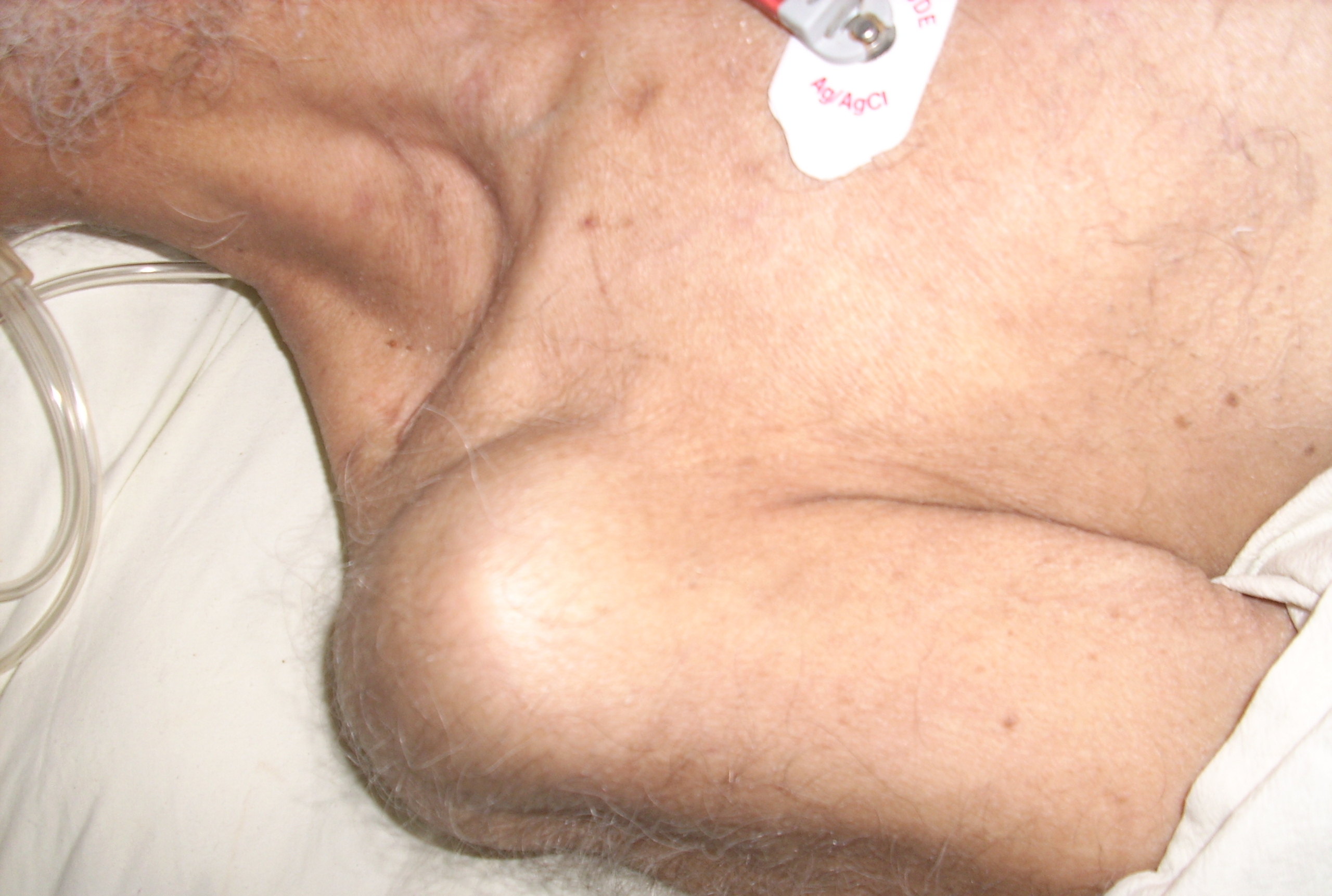
A dislocated shoulder is a condition in which the head of the humerus is detached from the shoulder joint. Symptoms include shoulder pain and instability. Complications may include a Bankart lesion, Hill-Sachs lesion, rotator cuff tear, or injury to the axillary nerve. A shoulder dislocation often occurs as a result of a fall onto an outstretched arm or onto the shoulder. Diagnosis is typically based on symptoms and confirmed by X-rays. They are classified as anterior, posterior, inferior, and superior with most being anterior. Treatment is by shoulder reduction which may be accomplished by a number of techniques. These include traction-countertraction, external rotation, scapular manipulation, and the Stimson technique. After reduction X-rays are recommended for verification. The arm may then be placed in a sling for a few weeks. Surgery may be recommended in those with recurrent dislocations. Not all patients require surgery following a shoulder dislocation. There is m ... [...More Info...] [...Related Items...] OR: [Wikipedia] [Google] [Baidu] |
Femoroacetabular Impingement
Femoroacetabular impingement (FAI) is a condition involving one or more anatomical abnormalities of the hip joint, which is a ball and socket joint. It is a common cause of hip pain and discomfort in young and middle-aged adults. It occurs when the ball shaped femoral head contacts the acetabulum abnormally or does not permit a normal range of motion in the acetabular socket. Damage can occur to the articular cartilage, or labral cartilage (soft tissue, ring-shaped bumper of the socket), or both. The condition may be symptomatic or asymptomatic. It may cause osteoarthritis of the hip. Treatment options range from conservative management to surgery. Signs and symptoms Pain is the most common complaint in those with FAI. It is experienced in a number of areas, making the diagnosis challenging, but commonly occurs in the groin, upper buttock/lower back, the buttock or beneath the buttock, side of the affected hip and posterior upper leg. Onset of symptoms has been reported to pres ... [...More Info...] [...Related Items...] OR: [Wikipedia] [Google] [Baidu] |
Rotator Cuff Tear
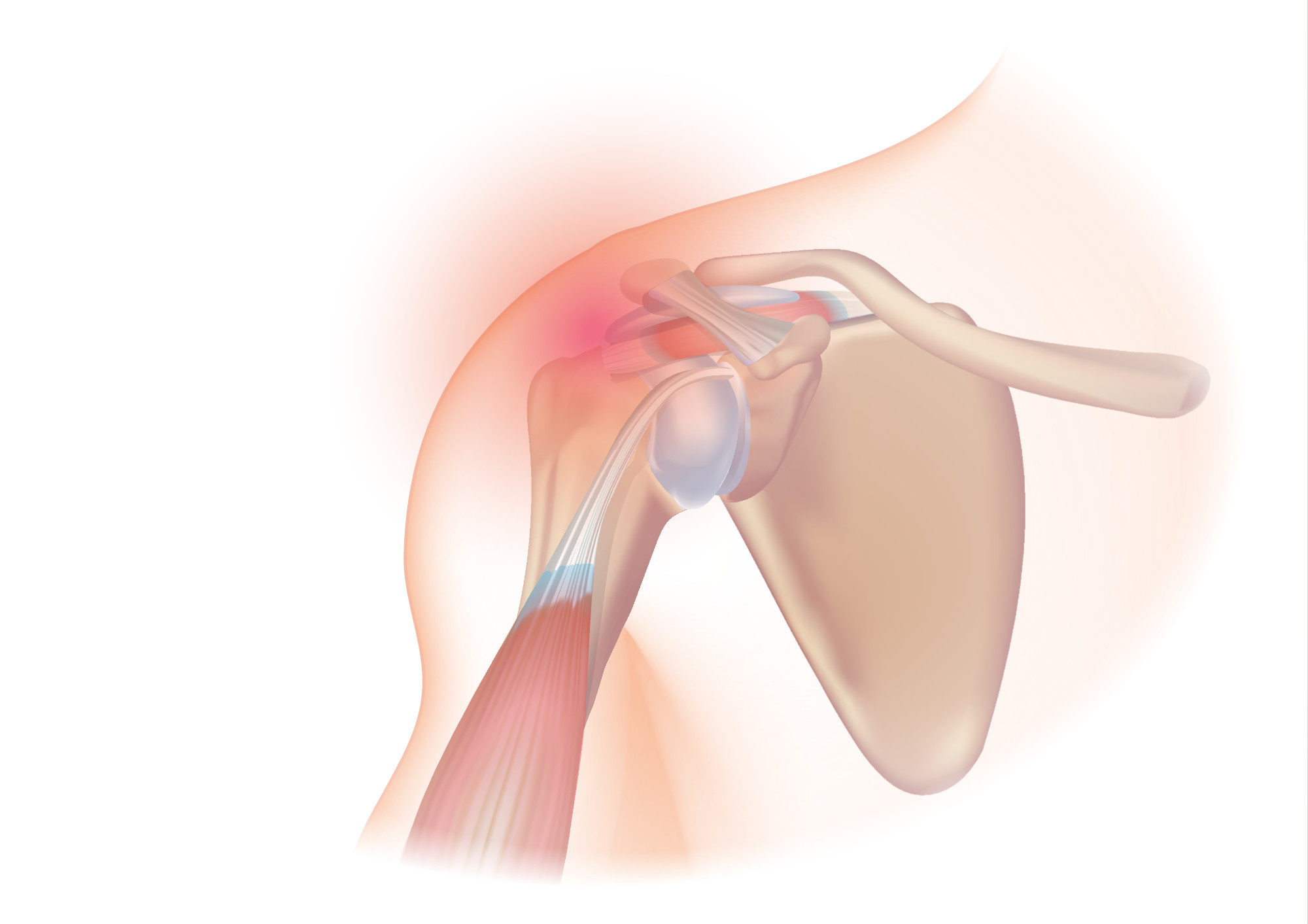
A rotator cuff tear is an injury where one or more of the tendons or muscles of the rotator cuff of the shoulder get torn. Symptoms may include shoulder pain, which is often worse with movement, limited range of motion, or weakness. This may limit people's ability to brush their hair or put on clothing. Clicking may also occur with movement of the arm. Tears may occur as the result of a sudden force or gradually over time. Risk factors include certain repetitive activities, smoking, and a family history of the condition. Diagnosis is based on symptoms, examination, and medical imaging. The rotator cuff is made up of the supraspinatus, infraspinatus, teres minor, and subscapularis. The supraspinatus is the most commonly affected. Treatment may include pain medication such as NSAIDs and specific exercises. It is recommended that people who are unable to raise their arm above 90 degrees after 2 weeks should be further assessed. In severe cases surgery may be tried, however ben ... [...More Info...] [...Related Items...] OR: [Wikipedia] [Google] [Baidu] |
Tear Of Meniscus
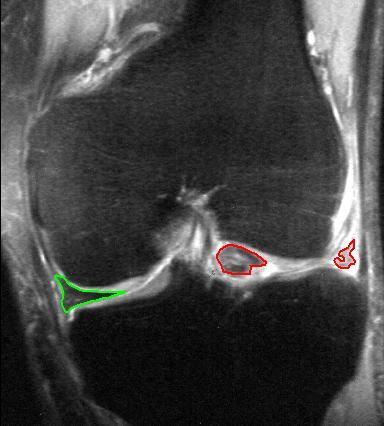
A tear of a meniscus is a rupturing of one or more of the fibrocartilage strips in the knee called menisci. When doctors and patients refer to "torn cartilage" in the knee, they actually may be referring to an injury to a meniscus at the top of one of the tibiae. Menisci can be torn during innocuous activities such as walking or squatting. They can also be torn by traumatic force encountered in sports or other forms of physical exertion. The traumatic action is most often a twisting movement at the knee while the leg is bent. In older adults, the meniscus can be damaged following prolonged 'wear and tear'. Especially acute injuries (typically in younger, more active patients) can lead to displaced tears which can cause mechanical symptoms such as clicking, catching, or locking during motion of the joint. The joint will be in pain when in use, but when there is no load, the pain goes away. A tear of the medial meniscus can occur as part of the unhappy triad, together with a tear ... [...More Info...] [...Related Items...] OR: [Wikipedia] [Google] [Baidu] |
Osteoarthritis
Osteoarthritis (OA) is a type of degenerative joint disease that results from breakdown of joint cartilage and underlying bone which affects 1 in 7 adults in the United States. It is believed to be the fourth leading cause of disability in the world. The most common symptoms are joint pain and stiffness. Usually the symptoms progress slowly over years. Initially they may occur only after exercise but can become constant over time. Other symptoms may include joint swelling, decreased range of motion, and, when the back is affected, weakness or numbness of the arms and legs. The most commonly involved joints are the two near the ends of the fingers and the joint at the base of the thumbs; the knee and hip joints; and the joints of the neck and lower back. Joints on one side of the body are often more affected than those on the other. The symptoms can interfere with work and normal daily activities. Unlike some other types of arthritis, only the joints, not internal organs, are af ... [...More Info...] [...Related Items...] OR: [Wikipedia] [Google] [Baidu] |
Anterior Cruciate Ligament
The anterior cruciate ligament (ACL) is one of a pair of cruciate ligaments (the other being the posterior cruciate ligament) in the human knee. The two ligaments are also called "cruciform" ligaments, as they are arranged in a crossed formation. In the quadruped stifle joint (analogous to the knee), based on its anatomical position, it is also referred to as the cranial cruciate ligament. The term cruciate translates to cross. This name is fitting because the ACL crosses the posterior cruciate ligament to form an “X”. It is composed of strong, fibrous material and assists in controlling excessive motion. This is done by limiting mobility of the joint. The anterior cruciate ligament is one of the four main ligaments of the knee, providing 85% of the restraining force to anterior tibial displacement at 30 and 90° of knee flexion. The ACL is the most injured ligament of the four located in the knee. Structure The ACL originates from deep within the notch of the distal fe ... [...More Info...] [...Related Items...] OR: [Wikipedia] [Google] [Baidu] |
Endoscope
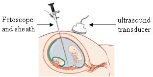
An endoscope is an inspection instrument composed of image sensor, optical lens, light source and mechanical device, which is used to look deep into the body by way of openings such as the mouth or anus. A typical endoscope applies several modern technologies including optics, ergonomics, precision mechanics, electronics, and software engineering. With an endoscope, it is possible to observe lesions that cannot be detected by X-ray, making it useful in medical diagnosis. Endoscopes use tubes which are only a few millimeters thick to transfer illumination in one direction and high-resolution images in real time in the other direction, resulting in minimally invasive surgeries. It is used to examine the internal organs like the throat or esophagus. Specialized instruments are named after their target organ. Examples include the cystoscope (bladder), nephroscope (kidney), bronchoscope (bronchus), arthroscope (joints) and colonoscope (colon), and laparoscope (abdomen or pelvis). They c ... [...More Info...] [...Related Items...] OR: [Wikipedia] [Google] [Baidu] |
Frozen Shoulder
Adhesive capsulitis, also known as frozen shoulder, is a condition associated with shoulder pain and stiffness. It is a common shoulder ailment that is marked by pain and a loss of range of motion, particularly in external rotation. There is a loss of the ability to move the shoulder, both voluntarily and by others, in multiple directions. The shoulder itself, however, does not generally hurt significantly when touched. Muscle loss around the shoulder may also occur. Onset is gradual over weeks to months. Complications can include fracture of the humerus or biceps tendon rupture. The cause in most cases is unknown. The condition can also occur after injury or surgery to the shoulder. Risk factors include diabetes and thyroid disease. The underlying mechanism involves inflammation and scarring. The diagnosis is generally based on a person's symptoms and a physical exam. The diagnosis may be supported by an MRI. Adhesive capsulitis has been linked to diabetes and hypothyroidism ... [...More Info...] [...Related Items...] OR: [Wikipedia] [Google] [Baidu] |


