|
Arcuate Line Of Ilium
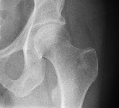
The arcuate line of the ilium is a smooth rounded border on the internal surface of the ilium. It is immediately inferior to the iliac fossa and Iliacus muscle. It forms part of the border of the pelvic inlet. In combination with the pectineal line, it comprises the iliopectineal line. The arcuate line marks the border between the body (''corpus'') and the wing (''ala'') of the ilium, and, running inferior, anterior, and medial from the auricular surface to the area corresponding to the acetabulum, it also indicates where weight is transferred from the sacroiliac joint to the hip joint In vertebrate anatomy, hip (or "coxa"Latin ''coxa'' was used by Celsus in the sense "hip", but by Pliny the Elder in the sense "hip bone" (Diab, p 77) in medical terminology) refers to either an anatomical region or a joint. The hip region is .... Additional images Arcuate line of ilium 02 animation.gif, Position of arcuate line of ilium. Shown in red. Arcuate line of ilium 03 animatio ... [...More Info...] [...Related Items...] OR: [Wikipedia] [Google] [Baidu] |
Ilium (bone)
The ilium () (plural ilia) is the uppermost and largest part of the hip bone, and appears in most vertebrates including mammals and birds, but not bony fish. All reptiles have an ilium except snakes, although some snake species have a tiny bone which is considered to be an ilium. The ilium of the human is divisible into two parts, the body and the wing; the separation is indicated on the top surface by a curved line, the arcuate line, and on the external surface by the margin of the acetabulum. The name comes from the Latin (''ile'', ''ilis''), meaning "groin" or "flank". Structure The ilium consists of the body and wing. Together with the ischium and pubis, to which the ilium is connected, these form the pelvic bone, with only a faint line indicating the place of union. The body ( la, corpus) forms less than two-fifths of the acetabulum; and also forms part of the acetabular fossa. The internal surface of the body is part of the wall of the lesser pelvis and gives or ... [...More Info...] [...Related Items...] OR: [Wikipedia] [Google] [Baidu] |
Iliac Fossa
The iliac fossa is a large, smooth, concave surface on the internal surface of the ilium (part of the three fused bones making the hip bone). Structure The iliac fossa is bounded above by the iliac crest, and below by the arcuate line. It is bordered in front and behind by the anterior and posterior borders of the ilium. The iliac fossa gives origin to the iliacus muscle. The obturator nerve passes around the iliac fossa. It is perforated at its inner part by a nutrient canal. Below it there is a smooth, rounded border, the arcuate line, which runs anterior, inferior, and medial. When the "left" or "right" adjective is used (e.g "right iliac fossa"), the iliac fossa usually means one of the inguinal regions of the nine regions of the abdomen The abdomen (colloquially called the belly, tummy, midriff, tucky or stomach) is the part of the body between the thorax (chest) and pelvis, in humans and in other vertebrates. The abdomen is the front part of the abdominal segmen ... [...More Info...] [...Related Items...] OR: [Wikipedia] [Google] [Baidu] |
Iliacus
The iliacus is a flat, triangular muscle which fills the iliac fossa. It forms the lateral portion of iliopsoas, providing flexion of the thigh and lower limb at the acetabulofemoral joint. Structure The iliacus arises from the iliac fossa on the interior side of the hip bone, and also from the region of the anterior inferior iliac spine (AIIS). It joins the psoas major to form the iliopsoas. It proceeds across the iliopubic eminence through the muscular lacuna to its insertion on the lesser trochanter of the femur. Its fibers are often inserted in front of those of the psoas major and extend distally over the lesser trochanter.Platzer (2004), p 234 Nerve supply The iliopsoas is innervated by the femoral nerve and direct branches from the lumbar plexus.''Thieme Atlas of Anatomy'' (2006), p 422 Function In open-chain exercises, as part of the iliopsoas, the iliacus is important for lifting (flexing) the femur forward (e.g. front scale). In closed-chain exercises, the iliopsoas b ... [...More Info...] [...Related Items...] OR: [Wikipedia] [Google] [Baidu] |
Pelvic Inlet
The pelvic inlet or superior aperture of the pelvis is a planar surface which defines the boundary between the pelvic cavity and the abdominal cavity (or, according to some authors, between two parts of the pelvic cavity, called lesser pelvis and greater pelvis). It is a major target of measurements of pelvimetry. Its position and orientation relative to the skeleton of the pelvis is anatomically defined by its edge, the pelvic brim. The pelvic brim is an approximately apple-shaped line passing through the prominence of the sacrum, the arcuate and pectineal lines, and the upper margin of the pubic symphysis. Occasionally, the terms pelvic inlet and pelvic brim are used interchangeably. Boundaries The edge of the pelvic inlet (pelvic brim) is formed as follows: Diameters The diameters or conjugates of the pelvis are measured at the pelvic inlet and outlet and as oblique diameters. Two diameters may be measured from the outside of the body using a pelvimeter Addition ... [...More Info...] [...Related Items...] OR: [Wikipedia] [Google] [Baidu] |
Pectineal Line (pubis)
The pectineal line of the pubis (also pecten pubis) is a ridge on the superior ramus of the pubic bone. It forms part of the pelvic brim. Lying across from the pectineal line are fibers of the pectineal ligament, and the proximal origin of the pectineus muscle. In combination with the arcuate line, it makes the iliopectineal line. References External links * () {{Authority control Bones of the pelvis Pubis (bone) ... [...More Info...] [...Related Items...] OR: [Wikipedia] [Google] [Baidu] |
Iliopectineal Line
The iliopectineal line is the border of the iliopubic eminence. It can be defined as a compound structure of the arcuate line (from the ilium) and pectineal line (from the pubis). With the sacral promontory, it makes up the linea terminalis The linea terminalis or innominate line consists of the pubic crest, pectineal line (pecten pubis), the arcuate line, the sacral ala, and the sacral promontory. It is the pelvic brim, which is the edge of the pelvic inlet. The pelvic inlet is ty .... The Iliopectineal line divides the pelvis into the pelvis major (false pelvis) above and the pelvis minor (true pelvis) below. References External links * http://ect.downstate.edu/courseware/haonline/labs/l43/st0217.htm {{Pelvis Bones of the pelvis ... [...More Info...] [...Related Items...] OR: [Wikipedia] [Google] [Baidu] |
Wing Of Ilium
The wing (ala) of ilium is the large expanded portion of the ilium, the bone which bounds the greater pelvis laterally. It presents for examination two surfaces—an external and an internal—a crest, and two borders—an anterior and a posterior. External surface The external surface, known as the dorsum ossis ilium, is directed backward and lateralward behind, and downward and lateralward in front. It is smooth, convex in front, deeply concave behind; bounded above by the crest, below by the upper border of the acetabulum, in front and behind by the anterior and posterior borders. This surface is crossed in an arched direction by three lines—the posterior, anterior, and inferior gluteal lines. The ''posterior gluteal line'' (superior curved line), the shortest of the three, begins at the crest, about 5 cm in front of its posterior extremity; it is at first distinctly marked, but as it passes downward to the upper part of the greater sciatic notch, where it ends, it ... [...More Info...] [...Related Items...] OR: [Wikipedia] [Google] [Baidu] |
Acetabulum
The acetabulum (), also called the cotyloid cavity, is a concave surface of the pelvis. The head of the femur meets with the pelvis at the acetabulum, forming the hip joint. Structure There are three bones of the ''os coxae'' (hip bone) that come together to form the ''acetabulum''. Contributing a little more than two-fifths of the structure is the ischium, which provides lower and side boundaries to the acetabulum. The ilium forms the upper boundary, providing a little less than two-fifths of the structure of the acetabulum. The rest is formed by the pubis, near the midline. It is bounded by a prominent uneven rim, which is thick and strong above, and serves for the attachment of the acetabular labrum, which reduces its opening, and deepens the surface for formation of the hip joint. At the lower part of the ''acetabulum'' is the acetabular notch, which is continuous with a circular depression, the acetabular fossa, at the bottom of the cavity of the ''acetabulum''. The ... [...More Info...] [...Related Items...] OR: [Wikipedia] [Google] [Baidu] |
Sacroiliac Joint
The sacroiliac joint or SI joint (SIJ) is the joint between the sacrum and the ilium bones of the pelvis, which are connected by strong ligaments. In humans, the sacrum supports the spine and is supported in turn by an ilium on each side. The joint is strong, supporting the entire weight of the upper body. It is a synovial plane joint with irregular elevations and depressions that produce interlocking of the two bones. The human body has two sacroiliac joints, one on the left and one on the right, that often match each other but are highly variable from person to person. Structure Sacroiliac joints are paired C-shaped or L-shaped joints capable of a small amount of movement (2–18 degrees, which is debatable at this time) that are formed between the auricular surfaces of the sacrum and the ilium bones. However mostBogduk, Nicolai "Clinical and Radiological Anatomy of the Lumbar Spine" Elsevier Health Sciences, 2022, p. 172. agree that there are only slight movements occ ... [...More Info...] [...Related Items...] OR: [Wikipedia] [Google] [Baidu] |
Hip Joint
In vertebrate anatomy, hip (or "coxa"Latin ''coxa'' was used by Celsus in the sense "hip", but by Pliny the Elder in the sense "hip bone" (Diab, p 77) in medical terminology) refers to either an anatomical region or a joint. The hip region is located lateral and anterior to the gluteal region, inferior to the iliac crest, and overlying the greater trochanter of the femur, or "thigh bone". In adults, three of the bones of the pelvis have fused into the hip bone or acetabulum which forms part of the hip region. The hip joint, scientifically referred to as the acetabulofemoral joint (''art. coxae''), is the joint between the head of the femur and acetabulum of the pelvis and its primary function is to support the weight of the body in both static (e.g., standing) and dynamic (e.g., walking or running) postures. The hip joints have very important roles in retaining balance, and for maintaining the pelvic inclination angle. Pain of the hip may be the result of numerous cau ... [...More Info...] [...Related Items...] OR: [Wikipedia] [Google] [Baidu] |
Bones Of The Pelvis
A bone is a rigid organ that constitutes part of the skeleton in most vertebrate animals. Bones protect the various other organs of the body, produce red and white blood cells, store minerals, provide structure and support for the body, and enable mobility. Bones come in a variety of shapes and sizes and have complex internal and external structures. They are lightweight yet strong and hard and serve multiple functions. Bone tissue (osseous tissue), which is also called bone in the uncountable sense of that word, is hard tissue, a type of specialized connective tissue. It has a honeycomb-like matrix internally, which helps to give the bone rigidity. Bone tissue is made up of different types of bone cells. Osteoblasts and osteocytes are involved in the formation and mineralization of bone; osteoclasts are involved in the resorption of bone tissue. Modified (flattened) osteoblasts become the lining cells that form a protective layer on the bone surface. The mineralized m ... [...More Info...] [...Related Items...] OR: [Wikipedia] [Google] [Baidu] |


