|
Arachnoid Mater
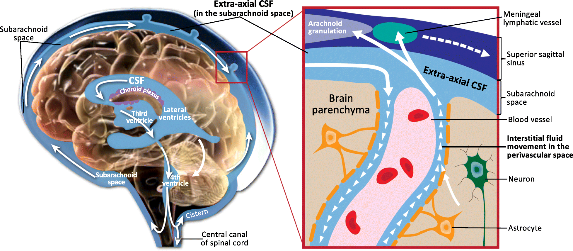
The arachnoid mater (or simply arachnoid) is one of the three meninges, the protective membranes that cover the brain and spinal cord. It is so named because of its resemblance to a spider web. The arachnoid mater is a derivative of the neural crest mesoectoderm in the embryo. Structure It is interposed between the two other meninges, the more superficial and much thicker dura mater and the deeper pia mater, from which it is separated by the subarachnoid space. The delicate arachnoid layer is not attached to the inside of the dura but against it and surrounds the brain and spinal cord. It does not line the brain down into its sulci (folds), as does the pia mater, with the exception of the longitudinal fissure, which divides the left and right cerebral hemispheres. Cerebrospinal fluid (CSF) flows under the arachnoid in the subarachnoid space, within a meshwork of trabeculae which span between the arachnoid and the pia. The arachnoid mater makes arachnoid villi, small protrusions ... [...More Info...] [...Related Items...] OR: [Wikipedia] [Google] [Baidu] |
Subarachnoid Space
In anatomy, the meninges (, ''singular:'' meninx ( or ), ) are the three membranes that envelop the brain and spinal cord. In mammals, the meninges are the dura mater, the arachnoid mater, and the pia mater. Cerebrospinal fluid is located in the subarachnoid space between the arachnoid mater and the pia mater. The primary function of the meninges is to protect the central nervous system. Structure Dura mater The dura mater ( la, tough mother) (also rarely called ''meninx fibrosa'' or ''pachymeninx'') is a thick, durable membrane, closest to the skull and vertebrae. The dura mater, the outermost part, is a loosely arranged, fibroelastic layer of cells, characterized by multiple interdigitating cell processes, no extracellular collagen, and significant extracellular spaces. The middle region is a mostly fibrous portion. It consists of two layers: the endosteal layer, which lies closest to the skull, and the inner meningeal layer, which lies closer to the brain. It contains large ... [...More Info...] [...Related Items...] OR: [Wikipedia] [Google] [Baidu] |
Arachnoid Trabeculae
The arachnoid trabeculae are delicate strands of connective tissue that loosely connect the two innermost layers of the meninges – the arachnoid mater and the pia mater. They are found within the subarachnoid space where cerebrospinal fluid is also found. Embryologically, the trabeculae are the remnants of the common precursor that forms both the arachnoid and pial layers of the meninges.Farr, Gary, MD"The Nervous System - Advanced Version / The Meninges", 24 Jun 2002. Retrieved 9 Sep 2010. See also *Subarachnoid space *Arachnoid mater *Trabeculae A trabecula (plural trabeculae, from Latin for "small beam") is a small, often microscopic, tissue element in the form of a small beam, strut or rod that supports or anchors a framework of parts within a body or organ. A trabecula generally has ... Notes {{Authority control Meninges ... [...More Info...] [...Related Items...] OR: [Wikipedia] [Google] [Baidu] |
Virchow-Robin Space
A perivascular space, also known as a Virchow–Robin space, is a fluid-filled space surrounding certain blood vessels in several organs, including the brain, potentially having an immunological function, but more broadly a dispersive role for neural and blood-derived messengers. The brain pia mater is reflected from the surface of the brain onto the surface of blood vessels in the subarachnoid space. In the brain, ''perivascular cuffs'' are regions of leukocyte aggregation in the perivascular spaces, usually found in patients with viral encephalitis. Perivascular spaces vary in dimension according to the type of blood vessel. In the brain where most capillaries have an imperceptible perivascular space, select structures of the brain, such as the circumventricular organs, are notable for having large perivascular spaces surrounding highly permeable capillaries, as observed by microscopy. The median eminence, a brain structure at the base of the hypothalamus, contains capillar ... [...More Info...] [...Related Items...] OR: [Wikipedia] [Google] [Baidu] |
Medulla Spinalis
The spinal cord is a long, thin, tubular structure made up of nervous tissue, which extends from the medulla oblongata in the brainstem to the lumbar region of the vertebral column (backbone). The backbone encloses the central canal of the spinal cord, which contains cerebrospinal fluid. The brain and spinal cord together make up the central nervous system (CNS). In humans, the spinal cord begins at the occipital bone, passing through the foramen magnum and then enters the spinal canal at the beginning of the cervical vertebrae. The spinal cord extends down to between the first and second lumbar vertebrae, where it ends. The enclosing bony vertebral column protects the relatively shorter spinal cord. It is around long in adult men and around long in adult women. The diameter of the spinal cord ranges from in the cervical and lumbar regions to in the thoracic area. The spinal cord functions primarily in the transmission of nerve signals from the motor cortex to the body, an ... [...More Info...] [...Related Items...] OR: [Wikipedia] [Google] [Baidu] |
Arachnoid Trabeculae
The arachnoid trabeculae are delicate strands of connective tissue that loosely connect the two innermost layers of the meninges – the arachnoid mater and the pia mater. They are found within the subarachnoid space where cerebrospinal fluid is also found. Embryologically, the trabeculae are the remnants of the common precursor that forms both the arachnoid and pial layers of the meninges.Farr, Gary, MD"The Nervous System - Advanced Version / The Meninges", 24 Jun 2002. Retrieved 9 Sep 2010. See also *Subarachnoid space *Arachnoid mater *Trabeculae A trabecula (plural trabeculae, from Latin for "small beam") is a small, often microscopic, tissue element in the form of a small beam, strut or rod that supports or anchors a framework of parts within a body or organ. A trabecula generally has ... Notes {{Authority control Meninges ... [...More Info...] [...Related Items...] OR: [Wikipedia] [Google] [Baidu] |
Arachne
Arachne (; from , cognate with Latin ) is the protagonist of a tale in Greek mythology known primarily from the version told by the Roman poet Ovid (43 BCE–17 CE), which is the earliest extant source for the story. In Book Six of his epic poem ''Metamorphoses'', Ovid recounts how the talented mortal Arachne, daughter of Idmon, challenged Athena, goddess of wisdom and crafts, to a weaving contest. When Athena could find no flaws in the tapestry Arachne had woven for the contest, the goddess became enraged and beat the girl with her shuttle. After Arachne hanged herself out of shame, she was transformed into a spider. The myth both provides an aetiology of spiders' web-spinning abilities and is a cautionary tale warning mortals not to place themselves on an equal level with the gods. Biography According to the myth as recounted by Ovid, Arachne was a Lydian maiden who was the daughter of Idmon of Colophon, who was a famous dyer in purple. She was credited to have in ... [...More Info...] [...Related Items...] OR: [Wikipedia] [Google] [Baidu] |
Greek Language
Greek ( el, label=Modern Greek, Ελληνικά, Elliniká, ; grc, Ἑλληνική, Hellēnikḗ) is an independent branch of the Indo-European family of languages, native to Greece, Cyprus, southern Italy (Calabria and Salento), southern Albania, and other regions of the Balkans, the Black Sea coast, Asia Minor, and the Eastern Mediterranean. It has the longest documented history of any Indo-European language, spanning at least 3,400 years of written records. Its writing system is the Greek alphabet, which has been used for approximately 2,800 years; previously, Greek was recorded in writing systems such as Linear B and the Cypriot syllabary. The alphabet arose from the Phoenician script and was in turn the basis of the Latin, Cyrillic, Armenian, Coptic, Gothic, and many other writing systems. The Greek language holds a very important place in the history of the Western world. Beginning with the epics of Homer, ancient Greek literature includes many works of lasting impo ... [...More Info...] [...Related Items...] OR: [Wikipedia] [Google] [Baidu] |
Choroid Plexus
The choroid plexus, or plica choroidea, is a plexus of cells that arises from the tela choroidea in each of the ventricles of the brain. Regions of the choroid plexus produce and secrete most of the cerebrospinal fluid (CSF) of the central nervous system. The choroid plexus consists of modified ependymal cells surrounding a core of capillaries and loose connective tissue. Multiple cilia on the ependymal cells move to circulate the cerebrospinal fluid. Structure Location There is a choroid plexus in each of the four ventricles. In the lateral ventricles it is found in the body, and continued in an enlarged amount in the atrium. There is no choroid plexus in the anterior horn. In the third ventricle there is a small amount in the roof that is continuous with that in the body, via the interventricular foramina, the channels that connect the lateral ventricles with the third ventricle. A choroid plexus is in part of the roof of the fourth ventricle. Microanatomy The chor ... [...More Info...] [...Related Items...] OR: [Wikipedia] [Google] [Baidu] |
Tela Choroidea
The tela choroidea (or tela chorioidea) is a region of meningeal pia mater that adheres to the underlying ependyma, and gives rise to the choroid plexus in each of the brain’s four ventricles. ''Tela'' is Latin for ''woven'' and is used to describe a web-like membrane or layer. The tela choroidea is a very thin part of the loose connective tissue of pia mater overlying and closely adhering to the ependyma. It has a rich blood supply. The ependyma and vascular pia mater – the tela choroidea, form regions of minute projections known as a choroid plexus that projects into each ventricle. The choroid plexus produces most of the cerebrospinal fluid of the central nervous system that circulates through the ventricles of the brain, the central canal of the spinal cord, and the subarachnoid space. The tela choroidea in the ventricles forms from different parts of the roof plate in the development of the embryo. Structure In the lateral ventricles the tela choroidea–a double-layere ... [...More Info...] [...Related Items...] OR: [Wikipedia] [Google] [Baidu] |
Leptomeninges
In anatomy, the meninges (, ''singular:'' meninx ( or ), ) are the three membranes that envelop the brain and spinal cord. In mammals, the meninges are the dura mater, the arachnoid mater, and the pia mater. Cerebrospinal fluid is located in the subarachnoid space between the arachnoid mater and the pia mater. The primary function of the meninges is to protect the central nervous system. Structure Dura mater The dura mater ( la, tough mother) (also rarely called ''meninx fibrosa'' or ''pachymeninx'') is a thick, durable membrane, closest to the skull and vertebrae. The dura mater, the outermost part, is a loosely arranged, fibroelastic layer of cells, characterized by multiple interdigitating cell processes, no extracellular collagen, and significant extracellular spaces. The middle region is a mostly fibrous portion. It consists of two layers: the endosteal layer, which lies closest to the skull, and the inner meningeal layer, which lies closer to the brain. It contains large ... [...More Info...] [...Related Items...] OR: [Wikipedia] [Google] [Baidu] |
Filum Terminale
The filum terminale ("terminal thread") is a delicate strand of fibrous tissue, about 20 cm in length, proceeding downward from the apex of the conus medullaris. It is one of the modifications of pia mater. It gives longitudinal support to the spinal cord and consists of two parts: * The upper part, or filum terminale internum, is about 15 cm long and reaches as far as the lower border of the second sacral vertebra. It is continuous above with the pia mater and contained within a tubular sheath of the dura mater. In addition, it is surrounded by the nerves forming the cauda equina, from which it can be easily recognized by its bluish-white color. * The lower part, or filum terminale externum, closely adheres to the dura mater. It extends downward from the apex of the tubular sheath and is attached to the back of the first segment of the coccyx in a structure sometimes referred to as the ''coccygeal ligament''. The most inferior of the spinal nerves, the coccygeal nerve l ... [...More Info...] [...Related Items...] OR: [Wikipedia] [Google] [Baidu] |
Dural Venous Sinus
The dural venous sinuses (also called dural sinuses, cerebral sinuses, or cranial sinuses) are venous channels found between the endosteal and meningeal layers of dura mater in the brain. They receive blood from the cerebral veins, receive cerebrospinal fluid (CSF) from the subarachnoid space via arachnoid granulations, and mainly empty into the internal jugular vein. Venous sinuses Structure The walls of the dural venous sinuses are composed of dura mater lined with endothelium, a specialized layer of flattened cells found in blood vessels. They differ from other blood vessels in that they lack a full set of vessel layers (e.g. tunica media) characteristic of arteries and veins. It also lacks valves (in veins; with exception of materno-fetal blood circulation i.e. placental artery and pulmonary arteries both of which carry deoxygenated blood). Clinical relevance The sinuses can be injured by trauma in which damage to the dura mater, may result in blood clot formation (throm ... [...More Info...] [...Related Items...] OR: [Wikipedia] [Google] [Baidu] |


.jpg)


