|
Anterolateral Ligament
The anterolateral ligament (ALL) is a ligament on the lateral aspect of the human knee, anterior to the fibular collateral ligament. Perhaps the earliest account of the ALL was written by French surgeon Paul Segond in 1879, in which he described a ligamentous structure between the lateral femur and tibia. Claes and Bellemans (2013) found that the ALL originates at the lateral epicondyle of the femur, and inserts at the anterolateral aspect of the proximal tibia The tibia (; ), also known as the shinbone or shankbone, is the larger, stronger, and anterior (frontal) of the two bones in the leg below the knee in vertebrates (the other being the fibula, behind and to the outside of the tibia); it connects .... However, Vincent et al. (2012) reported the origin to be the lateral femoral condyle. Clinical relevance The ALL, which has been suggested to occur in 33-97% of the human population, seems to stabilize medial rotation of the knee. However, the anterior cruciate ligame ... [...More Info...] [...Related Items...] OR: [Wikipedia] [Google] [Baidu] |
Lateral Epicondyle Of The Femur
The lateral epicondyle of the femur, smaller and less prominent than the medial epicondyle, gives attachment to the fibular collateral ligament of the knee-joint. Directly below it is a small depression from which a smooth well-marked groove curves obliquely upward and backward to the posterior extremity of the condyle A condyle (; in Bones of the lower limb Femur {{musculoskeletal-stub ... [...More Info...] [...Related Items...] OR: [Wikipedia] [Google] [Baidu] |
Avulsion Injury
In medicine, an avulsion is an injury in which a body structure is torn off by either trauma or surgery (from the Latin ''avellere'', meaning "to tear off"). The term most commonly refers to a surface trauma where all layers of the skin have been torn away, exposing the underlying structures (i.e., subcutaneous tissue, muscle, tendons, or bone). This is similar to an abrasion but more severe, as body parts such as an eyelid or an ear can be partially or fully detached from the body. Skin avulsions The most common avulsion injury, skin avulsion often occurs during motor vehicle collisions. The severity of avulsion ranges from skin flaps (minor) to degloving (moderate) and amputation of a finger or limb (severe). Suprafascial avulsions are those in which the depth of the removed skin reaches the subcutaneous tissue layer, while subfascial avulsions extend deeper than the subcutaneous layer.Jeng, S.F., & Wei, F.C. (1997, May). Classification and reconstructive options in foot plant ... [...More Info...] [...Related Items...] OR: [Wikipedia] [Google] [Baidu] |
Segond Fracture
The Segond fracture is a type of avulsion fracture (soft tissue structures pulling off fragments of their bony attachment) from the lateral tibial plateau of the knee, immediately below the articular surface of the tibia (see photo). Clinical significance Because of the high rate of associated ligamentous and meniscal injury, the presence of a Segond or reverse Segond fracture requires that these other pathologies must be specifically ruled out. Or more pragmatically, the orthopaedic team assume that the presence of Segond fracture equals ACL rupture, as the correlation is close to 100%. Increasingly, reconstruction of the ACL is combined with reconstruction of the anterolateral ligament when this associated pathology is present. The long-term benefit of an associated Anterolateral Ligament (ALL) reconstruction at the time of ACL surgery is not known. Attention and research is also being paid to a Lateral extra-articular Tenodesis (LET) instead of ALL. Diagnosis Segond and re ... [...More Info...] [...Related Items...] OR: [Wikipedia] [Google] [Baidu] |
Anterior Cruciate Ligament Injury
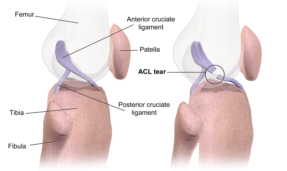
An anterior cruciate ligament injury occurs when the anterior cruciate ligament (ACL) is either stretched, partially torn, or completely torn. The most common injury is a complete tear. Symptoms include pain, an audible cracking sound during injury, instability of the knee, and joint swelling. Swelling generally appears within a couple of hours. In approximately 50% of cases, other structures of the knee such as surrounding ligaments, cartilage, or meniscus are damaged. The underlying mechanism often involves a rapid change in direction, sudden stop, landing after a jump, or direct contact to the knee. It is more common in athletes, particularly those who participate in alpine skiing, football (soccer), netball, American football, or basketball. Diagnosis is typically made by physical examination and is sometimes supported by magnetic resonance imaging (MRI). Physical examination will often show tenderness around the knee joint, reduced range of motion of the knee, and increase ... [...More Info...] [...Related Items...] OR: [Wikipedia] [Google] [Baidu] |
Medial Rotation
Motion, the process of movement, is described using specific anatomical terms. Motion includes movement of organs, joints, limbs, and specific sections of the body. The terminology used describes this motion according to its direction relative to the anatomical position of the body parts involved. Anatomists and others use a unified set of terms to describe most of the movements, although other, more specialized terms are necessary for describing unique movements such as those of the hands, feet, and eyes. In general, motion is classified according to the anatomical plane it occurs in. ''Flexion'' and ''extension'' are examples of ''angular'' motions, in which two axes of a joint are brought closer together or moved further apart. ''Rotational'' motion may occur at other joints, for example the shoulder, and are described as ''internal'' or ''external''. Other terms, such as ''elevation'' and ''depression'', describe movement above or below the horizontal plane. Many anatomica ... [...More Info...] [...Related Items...] OR: [Wikipedia] [Google] [Baidu] |
Lateral Femoral Condyle
The lateral condyle is one of the two projections on the lower extremity of the femur. The other one is the medial condyle. The lateral condyle is the more prominent and is broader both in its front-to-back and transverse diameters. Clinical significance The most common injury to the lateral femoral condyle is an osteochondral fracture combined with a patellar dislocation. The osteochondral fracture occurs on the weight-bearing portion of the lateral condyle. Typically, the condyle will fracture (and the patella may dislocate) as a result of severe impaction from activities such as downhill skiing and parachuting. Open reduction and internal fixation Internal fixation is an operation in orthopedics that involves the surgical implementation of implants for the purpose of repairing a bone, a concept that dates to the mid-nineteenth century and was made applicable for routine treatment in the m ... surgery is typically used to repair an osteochondral fracture. For a Type B1 parti ... [...More Info...] [...Related Items...] OR: [Wikipedia] [Google] [Baidu] |
Insertion (anatomy)
Anatomical terminology is used to uniquely describe aspects of skeletal muscle, cardiac muscle, and smooth muscle such as their actions, structure, size, and location. Types There are three types of muscle tissue in the body: skeletal, smooth, and cardiac. Skeletal muscle Skeletal muscle, or "voluntary muscle", is a striated muscle tissue that primarily joins to bone with tendons. Skeletal muscle enables movement of bones, and maintains posture. The widest part of a muscle that pulls on the tendons is known as the belly. Muscle slip A muscle slip is a slip of muscle that can either be an anatomical variant, or a branching of a muscle as in rib connections of the serratus anterior muscle. Smooth muscle Smooth muscle is involuntary and found in parts of the body where it conveys action without conscious intent. The majority of this type of muscle tissue is found in the digestive and urinary systems where it acts by propelling forward food, chyme, and feces in the forme ... [...More Info...] [...Related Items...] OR: [Wikipedia] [Google] [Baidu] |
Origin (anatomy)
Anatomical terminology is used to uniquely describe aspects of skeletal muscle, cardiac muscle, and smooth muscle such as their actions, structure, size, and location. Types There are three types of muscle tissue in the body: skeletal, smooth, and cardiac. Skeletal muscle Skeletal muscle, or "voluntary muscle", is a striated muscle tissue that primarily joins to bone with tendons. Skeletal muscle enables movement of bones, and maintains posture. The widest part of a muscle that pulls on the tendons is known as the belly. Muscle slip A muscle slip is a slip of muscle that can either be an anatomical variant, or a branching of a muscle as in rib connections of the serratus anterior muscle. Smooth muscle Smooth muscle is involuntary and found in parts of the body where it conveys action without conscious intent. The majority of this type of muscle tissue is found in the digestive and urinary systems where it acts by propelling forward food, chyme, and feces in the forme ... [...More Info...] [...Related Items...] OR: [Wikipedia] [Google] [Baidu] |
Tibia
The tibia (; ), also known as the shinbone or shankbone, is the larger, stronger, and anterior (frontal) of the two bones in the leg below the knee in vertebrates (the other being the fibula, behind and to the outside of the tibia); it connects the knee with the ankle. The tibia is found on the medial side of the leg next to the fibula and closer to the median plane. The tibia is connected to the fibula by the interosseous membrane of leg, forming a type of fibrous joint called a syndesmosis with very little movement. The tibia is named for the flute ''tibia''. It is the second largest bone in the human body, after the femur. The leg bones are the strongest long bones as they support the rest of the body. Structure In human anatomy, the tibia is the second largest bone next to the femur. As in other vertebrates the tibia is one of two bones in the lower leg, the other being the fibula, and is a component of the knee and ankle joints. The ossification or formation of the bone ... [...More Info...] [...Related Items...] OR: [Wikipedia] [Google] [Baidu] |
Johan Bellemans
Johan Bellemans is a Belgian former Olympic sailor and physician who became known for the announcement of "discovery" of the anterolateral ligament, already discovered by French doctor Paul Segond in 1879 . He discovered it along with Steven Claes in 2013, after a 134-year period of study for a missing part. In 2002 he also discovered the reason for limited flexion that is frequently noted after knee replacement surgery, and the concept of posterior condylar offset was originated by him. In 2014 he introduced the theory of constitutional alignment, which fundamentally changed modern thinking in knee surgery. He also was influential in the development of the fourth generation artificial knees, and developed the bicruciate stabilised Journey Knee system. Currently he works as an orthopedic surgeon in a regional hospital in Genk, Belgium after his highly mediatized resigning in both Katholieke Universiteit Leuven and UZ Leuven. In 1992 he represented Belgium at the 1992 Summer ... [...More Info...] [...Related Items...] OR: [Wikipedia] [Google] [Baidu] |
Paul Segond
Paul Ferdinand Segond (8 May 1851 in Paris – 27 October 1912 in Paris) was a French surgeon who was a founder of obstetrics and the teaching of gynaecology in Paris. He was also an expert on the knee and described the eponymous Segond fracture. Biography Paul Segond was born in Paris, the son of anatomist Louis-Auguste Segond (1819–1908). He studied medicine in Paris, becoming an intern in 1875, having already published a letter on "weight of newborns" in the ''Annales de gynécologie''. He became prosector at the Faculté de médecine de Paris of the University of Paris in 1878. He qualified docteur en médecine in 1880, with his thesis on ''Abcès chauds de la prostate et le phlegmon périprostatique'' (hot abscesses of the prostate and periprostatic phlegmon) being honoured by the Société de Chirurgie and French Academy of Sciences. He became an associate professor of surgery in 1883, and was made ''chef de clinique'' at Pitié-Salpêtrière Hospital alo ... [...More Info...] [...Related Items...] OR: [Wikipedia] [Google] [Baidu] |


