|
Anterior Ethmoidal Nerve
The anterior ethmoidal nerve is a nerve of the nose. It is a branch of the nasociliary nerve, itself a branch of the ophthalmic nerve (V1). It provides sensory innervation to some structures around the nasal cavity. Structure The anterior ethmoidal nerve is a terminal branch of the nasociliary nerve, a branch of the ophthalmic nerve (CN V1), itself a branch of the trigeminal nerve (CN V). It branches near the medial wall of the orbit. The anterior ethmoidal nerve arises only after the nasociliary has given off its four branches (the ramus communicans to the ciliary ganglion, the long ciliary nerves, the infratrochlear nerve, and the posterior ethmoidal nerve). It travels through the anterior ethmoidal foramen to reach the anterior cranial fossa. It then moves forward and passes through the cribriform plate to enter the nasal cavity. It gives off branches to the roof of the nasal cavity, and bifurcates into a lateral internal nasal branch and medial internal nasal branch. It send ... [...More Info...] [...Related Items...] OR: [Wikipedia] [Google] [Baidu] |
Nasociliary Nerve
The nasociliary nerve is a branch of the ophthalmic nerve, itself a branch of the trigeminal nerve (CN V). It is intermediate in size between the other two branches of the ophthalmic nerve, the frontal nerve and lacrimal nerve. Structure The nasociliary nerve enters the orbit via the superior orbital fissure, between the two heads of the lateral rectus muscle and between the superior and inferior rami of the oculomotor nerve. It passes across the optic nerve (CN II) and runs obliquely beneath the superior rectus muscle and superior oblique muscle to the medial wall of the orbital cavity. It passes through the anterior ethmoidal opening as the anterior ethmoidal nerve and enters the cranial cavity just below the cribriform plate of the ethmoid bone. It supplies branches to the mucous membrane of the nasal cavity and finally emerges between the inferior border of the nasal bone and the side nasal cartilages as the external nasal branch. Branches * posterior ethmoidal nerve * ante ... [...More Info...] [...Related Items...] OR: [Wikipedia] [Google] [Baidu] |
Anterior Ethmoidal Foramen
The anterior ethmoidal foramen is a small opening in the ethmoid bone in the skull. Lateral to either olfactory groove are the internal openings of the anterior and posterior ethmoidal foramina (or canals). The anterior ethmoidal foramen, situated about the middle of the lateral margin of the olfactory groove, transmits the anterior ethmoidal artery, vein and nerve. The anterior ethmoidal nerve, a branch of the nasociliary nerve, runs in a groove along the lateral edge of the cribriform plate In mammalian anatomy, the cribriform plate (Latin for lit. ''sieve-shaped''), horizontal lamina or lamina cribrosa is part of the ethmoid bone. It is received into the ethmoidal notch of the frontal bone and roofs in the nasal cavities. It supp ... to the above-mentioned slit-like opening . References External links * () (#5) Foramina of the skull {{musculoskeletal-stub ... [...More Info...] [...Related Items...] OR: [Wikipedia] [Google] [Baidu] |
Diving Reflex
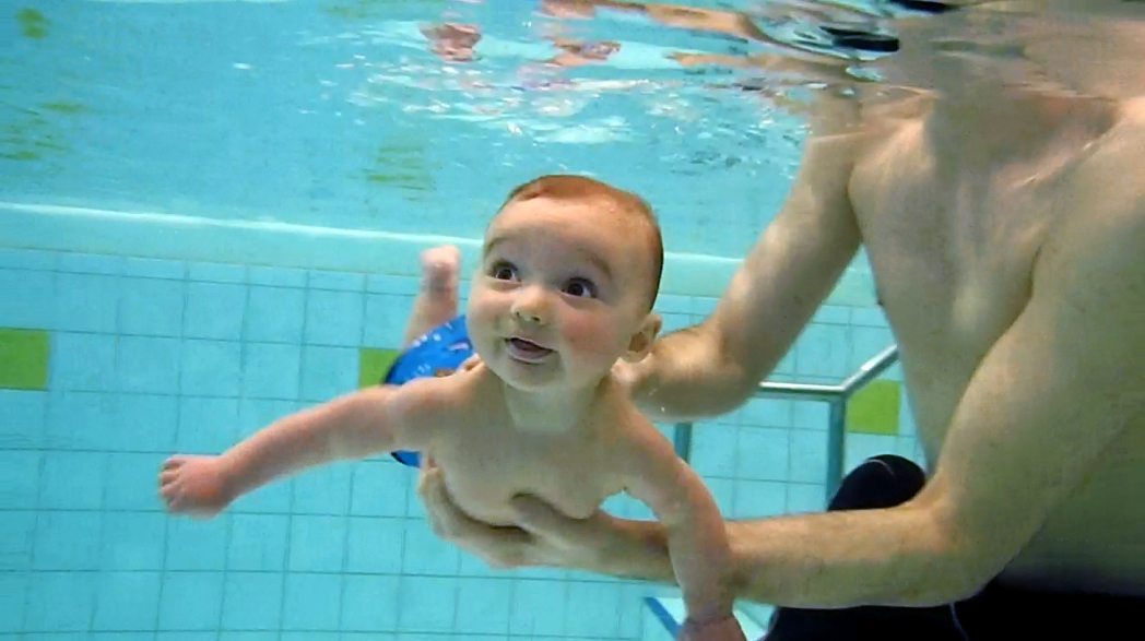
The diving reflex, also known as the diving response and mammalian diving reflex, is a set of physiological responses to immersion that overrides the basic homeostatic reflexes, and is found in all air-breathing vertebrates studied to date. It optimizes respiration by preferentially distributing oxygen stores to the heart and brain, enabling submersion for an extended time. The diving reflex is exhibited strongly in aquatic mammals, such as seals, otters, dolphins, and muskrats, and exists as a lesser response in other animals, including human babies up to 6 months old (see infant swimming), and diving birds, such as ducks and penguins. Adult humans generally exhibit a mild response, the dive-hunting Sama-Bajau people being a notable outlier. The diving reflex is triggered specifically by chilling and wetting the nostrils and face while breath-holding, and is sustained via neural processing originating in the carotid chemoreceptors. The most noticeable effects are on the cardiov ... [...More Info...] [...Related Items...] OR: [Wikipedia] [Google] [Baidu] |
Nasal Septum
The nasal septum () separates the left and right airways of the Human nose, nasal cavity, dividing the two nostrils. It is Depression (kinesiology), depressed by the depressor septi nasi muscle. Structure The fleshy external end of the nasal septum is called the Human nose#Cartilages, columella or columella nasi, and is made up of cartilage and soft tissue. The nasal septum contains bone and hyaline cartilage. It is normally about 2 mm thick. The nasal septum is composed of four structures: * Perpendicular plate of ethmoid bone * Vomer bone * Septal nasal cartilage * Maxillary bone (the crest) The lowest part of the septum is a narrow strip of bone that projects from the maxilla and the Palatine bone, palatine bones, and is the length of the septum. This strip of bone is called the maxillary crest; it articulates in front with the septal nasal cartilage, and at the back with the vomer. The maxillary crest is described in the anatomy of the nasal septum as having a maxill ... [...More Info...] [...Related Items...] OR: [Wikipedia] [Google] [Baidu] |
Elsevier
Elsevier () is a Dutch academic publishing company specializing in scientific, technical, and medical content. Its products include journals such as ''The Lancet'', ''Cell'', the ScienceDirect collection of electronic journals, '' Trends'', the '' Current Opinion'' series, the online citation database Scopus, the SciVal tool for measuring research performance, the ClinicalKey search engine for clinicians, and the ClinicalPath evidence-based cancer care service. Elsevier's products and services also include digital tools for data management, instruction, research analytics and assessment. Elsevier is part of the RELX Group (known until 2015 as Reed Elsevier), a publicly traded company. According to RELX reports, in 2021 Elsevier published more than 600,000 articles annually in over 2,700 journals; as of 2018 its archives contained over 17 million documents and 40,000 e-books, with over one billion annual downloads. Researchers have criticized Elsevier for its high profit marg ... [...More Info...] [...Related Items...] OR: [Wikipedia] [Google] [Baidu] |
Meninges
In anatomy, the meninges (, ''singular:'' meninx ( or ), ) are the three membranes that envelop the brain and spinal cord. In mammals, the meninges are the dura mater, the arachnoid mater, and the pia mater. Cerebrospinal fluid is located in the subarachnoid space between the arachnoid mater and the pia mater. The primary function of the meninges is to protect the central nervous system. Structure Dura mater The dura mater ( la, tough mother) (also rarely called ''meninx fibrosa'' or ''pachymeninx'') is a thick, durable membrane, closest to the Human skull, skull and vertebrae. The dura mater, the outermost part, is a loosely arranged, fibroelastic layer of cells, characterized by multiple interdigitating cell processes, no extracellular collagen, and significant extracellular spaces. The middle region is a mostly fibrous portion. It consists of two layers: the endosteal layer, which lies closest to the Calvaria (skull), skull, and the inner meningeal layer, which lies closer ... [...More Info...] [...Related Items...] OR: [Wikipedia] [Google] [Baidu] |
Ethmoid Sinus
The ethmoid sinuses or ethmoid air cells of the ethmoid bone are one of the four paired paranasal sinuses. The cells are variable in both size and number in the lateral mass of each of the ethmoid bones and cannot be palpated during an extraoral examination. They are divided into anterior and posterior groups. The ethmoid air cells are numerous thin-walled cavities situated in the ethmoidal labyrinth and completed by the frontal, maxilla, lacrimal, sphenoidal, and palatine bones. They lie between the upper parts of the nasal cavities and the orbits, and are separated from these cavities by thin bony lamellae. Groups of sinuses The groups of the ethmoidal air cells drain into the nasal meatuses.Otorhinolaryngology, Head and Neck Surgery, Anniko, Springer, 2010, page 188 * The posterior group the ''posterior ethmoidal sinus'' drains into the superior meatus above the middle nasal concha; sometimes one or more opens into the sphenoidal sinus. * The anterior group the ''anterior ethmo ... [...More Info...] [...Related Items...] OR: [Wikipedia] [Google] [Baidu] |
Cribriform Plate
In mammalian anatomy, the cribriform plate (Latin for lit. ''sieve-shaped''), horizontal lamina or lamina cribrosa is part of the ethmoid bone. It is received into the ethmoidal notch of the frontal bone and roofs in the nasal cavities. It supports the olfactory bulb, and is perforated by olfactory foramina for the passage of the olfactory nerves to the roof of the nasal cavity to convey smell to the brain. The foramina at the medial part of the groove allow the passage of the nerves to the upper part of the nasal septum while the foramina at the lateral part transmit the nerves to the superior nasal concha. A fractured cribriform plate can result in olfactory dysfunction, septal hematoma, cerebrospinal fluid rhinorrhoea (CSF rhinorrhoea), and possibly infection which can lead to meningitis. CSF rhinorrhoea (clear fluid leaking from the nose) is very serious and considered a medical emergency. Aging can cause the openings in the cribriform plate to close, pinching olfactor ... [...More Info...] [...Related Items...] OR: [Wikipedia] [Google] [Baidu] |
Anterior Cranial Fossa
The anterior cranial fossa is a depression in the floor of the cranial base which houses the projecting frontal lobes of the brain. It is formed by the orbital part of frontal bone, orbital plates of the Frontal bone, frontal, the cribriform plate of the ethmoid, and the small wings and front part of the body of the Sphenoid bone, sphenoid; it is limited behind by the posterior borders of the small wings of the sphenoid and by the anterior margin of the chiasmatic groove. The lesser wings of the sphenoid separate the anterior and middle cranial fossa, middle fossae. Structure It is traversed by the frontoethmoidal, sphenoethmoidal, and sphenofrontal sutures. Its lateral portions roof in the orbital cavities and support the frontal lobes of the cerebrum; they are convex and marked by depressions for the brain convolutions, and grooves for branches of the meningeal vessels. The central portion corresponds with the roof of the nasal cavity, and is markedly depressed on either side o ... [...More Info...] [...Related Items...] OR: [Wikipedia] [Google] [Baidu] |
Posterior Ethmoidal Nerve
The posterior ethmoidal nerve is a nerve of the orbit around the eye. It is a branch of the nasociliary nerve from the ophthalmic nerve (CN V1). It supplies sensation to the sphenoid sinus, the ethmoid sinus, and part of the dura mater in the anterior cranial fossa. Structure The posterior ethmoidal nerve is a branch of the nasociliary nerve, itself a branch of the ophthalmic nerve (CN V1), itself a branch of the trigeminal nerve (CN V). It passes through the posterior ethmoidal foramen, with the posterior ethmoidal artery. It gives branches to the sphenoid sinus and the ethmoid sinus. It also gives a branch to supply part of the dura mater in the anterior cranial fossa. Variation The posterior ethmoidal nerve is absent in a significant proportion of people. This may be around 30%. Function The posterior ethmoidal nerve supplies sensation to the sphenoid sinus and the ethmoid sinus. It also supplies sensation to part of the dura mater in the anterior cranial fossa. Oth ... [...More Info...] [...Related Items...] OR: [Wikipedia] [Google] [Baidu] |
Nose
A nose is a protuberance in vertebrates that houses the nostrils, or nares, which receive and expel air for respiration alongside the mouth. Behind the nose are the olfactory mucosa and the sinuses. Behind the nasal cavity, air next passes through the pharynx, shared with the digestive system, and then into the rest of the respiratory system. In humans, the nose is located centrally on the face and serves as an alternative respiratory passage especially during suckling for infants. The protruding nose that completely separate from the mouth part is a characteristic found only in therian mammals. It has been theorized that this unique mammalian nose evolved from the anterior part of the upper jaw of the reptilian-like ancestors ( synapsids). Air treatment Acting as the first interface between the external environment and an animal's delicate internal lungs, a nose conditions incoming air, both as a function of thermal regulation and filtration during respiration, as well as ... [...More Info...] [...Related Items...] OR: [Wikipedia] [Google] [Baidu] |
Infratrochlear Nerve
The infratrochlear nerve is a branch of the nasociliary nerve, itself a branch of the ophthalmic nerve. Structure The nasociliary nerve terminates by bifurcating into the infratrochlear and the anterior ethmoidal nerves. The infratrochlear nerve travels anteriorly in the orbit along the upper border of the medial rectus muscle and underneath the trochlea of the superior oblique muscle. It exits the orbit medially and divides into small sensory branches. Function The infratrochlear nerve provides sensory innervation to the skin of the eye lids, the conjunctiva, lacrimal sac, lacrimal caruncle and the side of the nose above the medial canthus. Etymology The infratrochlear nerve is named after a structure it passes under. ''Infratrochlear'' means "below the trochlea Trochlea (Latin for pulley) is a term in anatomy. It refers to a grooved structure reminiscent of a pulley's wheel. Related to joints Most commonly, trochleae bear the articular surface of saddle joint, saddle ... [...More Info...] [...Related Items...] OR: [Wikipedia] [Google] [Baidu] |


