|
Anterior Branch Of Obturator Nerve
The anterior branch of the obturator nerve is a branch of the obturator nerve found in the pelvis and leg. It leaves the pelvis in front of the obturator externus and descends anterior to the adductor brevis, and posterior to the pectineus and adductor longus; at the lower border of the latter muscle it communicates with the anterior cutaneous and saphenous branches of the femoral nerve, forming a kind of plexus. It then descends upon the femoral artery, to which it is finally distributed. Near the obturator foramen the nerve gives off an articular branch to the hip joint. Behind the pectineus, it distributes branches to the adductor longus and gracilis, and usually to the adductor brevis, and in rare cases to the pectineus; it receives a communicating branch from the accessory obturator nerve In human anatomy, the accessory obturator nerve is an accessory nerve in the lumbar region present in about 29% of cases. It is of small size, and arises from the ventral divisions of ... [...More Info...] [...Related Items...] OR: [Wikipedia] [Google] [Baidu] |
Obturator Nerve
The obturator nerve in human anatomy arises from the ventral divisions of the second, third, and fourth lumbar nerves in the lumbar plexus; the branch from the third is the largest, while that from the second is often very small. Structure The obturator nerve originates from the anterior divisions of the L2, L3, and L4 spinal nerve roots. It descends through the fibers of the psoas major, and emerges from its medial border near the brim of the pelvis. It then passes behind the common iliac arteries, and on the lateral side of the internal iliac artery and vein, and runs along the lateral wall of the lesser pelvis, above and in front of the obturator vessels, to the upper part of the obturator foramen. Here it enters the thigh, through the obturator canal, and divides into an anterior and a posterior branch, which are separated at first by some of the fibers of the obturator externus, and lower down by the adductor brevis. An accessory obturator nerve may be present in approx ... [...More Info...] [...Related Items...] OR: [Wikipedia] [Google] [Baidu] |
Femoral Nerve
The femoral nerve is a nerve in the thigh that supplies skin on the upper thigh and inner leg, and the muscles that extend the knee. Structure The femoral nerve is the major nerve supplying the anterior compartment of the thigh. It is the largest branch of the lumbar plexus, and arises from the dorsal divisions of the ventral rami of the second, third, and fourth lumbar nerves (L2, L3, and L4). The nerve enters Scarpa's triangle by passing beneath the inguinal ligament, just lateral to the femoral artery. In the thigh, the nerve lies in a groove between iliacus muscle and psoas major muscle, outside the femoral sheath, and lateral to the femoral artery. After a short course of about 4 cm in the thigh, the nerve is divided into anterior and posterior divisions, separated by lateral femoral circumflex artery. The branches are shown below: Muscular branches * The nerve to the pectineus muscle arises immediately above the inguinal ligament from the medial side of the femoral n ... [...More Info...] [...Related Items...] OR: [Wikipedia] [Google] [Baidu] |
Gracilis Muscle
The gracilis muscle (; Latin for "slender") is the most superficial muscle on the medial side of the thigh. It is thin and flattened, broad above, narrow and tapering below. Structure It arises by a thin aponeurosis from the anterior margins of the lower half of the symphysis pubis and the upper half of the pubic arch. The muscle's fibers run vertically downward, ending in a rounded tendon. This tendon passes behind the medial condyle of the femur, curves around the medial condyle of the tibia where it becomes flattened, and inserts into the upper part of the medial surface of the body of the tibia, below the condyle. For this reason, the muscle is a lower limb adductor. At its insertion the tendon is situated immediately above that of the semitendinosus muscle, and its upper edge is overlapped by the tendon of the sartorius muscle, which it joins to form the pes anserinus. The pes anserinus is separated from the medial collateral ligament of the knee-joint by a bursa. A ... [...More Info...] [...Related Items...] OR: [Wikipedia] [Google] [Baidu] |
Longus Colli Muscle
The longus colli muscle (Latin for ''long muscle of the neck'') is a muscle of the human body. The longus colli is situated on the anterior surface of the vertebral column, between the atlas and the third thoracic vertebra. It is broad in the middle, narrow and pointed at either end, and consists of three portions, a superior oblique, an inferior oblique, and a vertical. * The ''superior oblique portion'' arises from the anterior tubercles of the transverse processes of the third, fourth, and fifth cervical vertebrae and, ascending obliquely with a medial inclination, is inserted by a narrow tendon into the tubercle on the anterior arch of the atlas. * The ''inferior oblique portion'', the smallest part of the muscle, arises from the front of the bodies of the first two or three thoracic vertebrae; and, ascending obliquely in a lateral direction, is inserted into the anterior tubercles of the transverse processes of the fifth and sixth cervical vertebrae. * The ''vertical portion'' ... [...More Info...] [...Related Items...] OR: [Wikipedia] [Google] [Baidu] |
Hip Joint
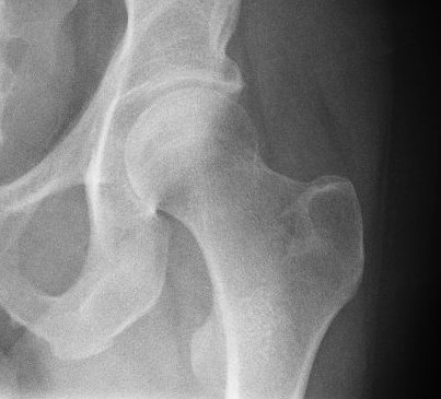
In vertebrate anatomy, hip (or "coxa"Latin ''coxa'' was used by Celsus in the sense "hip", but by Pliny the Elder in the sense "hip bone" (Diab, p 77) in medical terminology) refers to either an anatomical region or a joint. The hip region is located lateral and anterior to the gluteal region, inferior to the iliac crest, and overlying the greater trochanter of the femur, or "thigh bone". In adults, three of the bones of the pelvis have fused into the hip bone or acetabulum which forms part of the hip region. The hip joint, scientifically referred to as the acetabulofemoral joint (''art. coxae''), is the joint between the head of the femur and acetabulum of the pelvis and its primary function is to support the weight of the body in both static (e.g., standing) and dynamic (e.g., walking or running) postures. The hip joints have very important roles in retaining balance, and for maintaining the pelvic inclination angle. Pain of the hip may be the result of numerous cause ... [...More Info...] [...Related Items...] OR: [Wikipedia] [Google] [Baidu] |
Obturator Foramen
The obturator foramen (Latin foramen obturatum) is the large opening created by the ischium and pubis (bone), pubis bones of the pelvis through which nerves and blood vessels pass. Structure It is bounded by a thin, uneven margin, to which a strong membrane is attached, and presents, superiorly, a deep groove, the obturator groove, which runs from the pelvis obliquely medialward and downward. This groove is converted into the obturator canal by a ligamentous band, a specialized part of the obturator membrane, attached to two tubercles: * one, the posterior obturator tubercle, on the medial border of the ischium, just in front of the acetabular notch * the other, the anterior obturator tubercle, on the obturator crest of the superior pubic ramus, superior ramus of the pubis (bone), pubis Variation Reflecting the overall sex differences in human physiology, sex differences between male and female pelvises, the obturator foramina are oval in the male and wider and more triangular ... [...More Info...] [...Related Items...] OR: [Wikipedia] [Google] [Baidu] |
Femoral Artery
The femoral artery is a large artery in the thigh and the main arterial supply to the thigh and leg. The femoral artery gives off the deep femoral artery or profunda femoris artery and descends along the anteromedial part of the thigh in the femoral triangle. It enters and passes through the adductor canal, and becomes the popliteal artery as it passes through the adductor hiatus in the adductor magnus near the junction of the middle and distal thirds of the thigh. Structure The femoral artery enters the thigh from behind the inguinal ligament as the continuation of the external iliac artery. Here, it lies midway between the anterior superior iliac spine and the symphysis pubis (Mid-inguinal point). Segments In clinical parlance, the femoral artery has the following segments: *The common femoral artery (CFA) is the segment of the femoral artery between the inferior margin of the inguinal ligament and the branching point of the deep femoral artery/profunda femoris artery. Its ... [...More Info...] [...Related Items...] OR: [Wikipedia] [Google] [Baidu] |
Plexus
In neuroanatomy, a plexus (from the Latin term for "braid") is a branching network of vessels or nerves. The vessels may be blood vessels (veins, capillaries) or lymphatic vessels. The nerves are typically axons outside the central nervous system. The standard plural form in English is plexuses. Alternatively, the Latin plural plexūs may be used. Types Nerve plexuses The four primary nerve plexus A nerve plexus is a plexus (branching network) of intersecting nerves. A nerve plexus is composed of afferent and efferent fibers that arise from the merging of the anterior rami of spinal nerves and blood vessels. There are five spinal nerve ple ...es are the cervical plexus, brachial plexus, lumbar plexus, and the sacral plexus. Cardiac plexus Celiac plexus Renal plexus Venous plexus Choroid plexus The choroid plexus is a part of the central nervous system in the brain and consists of capillaries, brain ventricles, and ependymal cells. Invertebrates The plexus is the ch ... [...More Info...] [...Related Items...] OR: [Wikipedia] [Google] [Baidu] |
Saphenous Nerve
The saphenous nerve (long or internal saphenous nerve) is the largest cutaneous branch Cutaneous innervation refers to the area of the skin which is supplied by a specific cutaneous nerve. Dermatome (Anatomy), Dermatomes are similar; however, a dermatome only specifies the area served by a spinal nerve. In some cases, the dermatome i ... of the femoral nerve. It is a strictly sensory nerve, and has no motor function. Structure It is purely a sensory nerve. The saphenous nerve is the largest and terminal branch of the femoral nerve. Shortly after the femoral nerve passes under the inguinal ligament, it splits into anterior and posterior divisions by the passage of the lateral femoral circumflex artery (a branch of the profunda femoris artery). The posterior division then gives off the saphenous nerve as it converges with the femoral artery where it passes beneath the sartorius muscle. The saphenous nerve lies in front of the femoral artery, behind the aponeurotic covering of th ... [...More Info...] [...Related Items...] OR: [Wikipedia] [Google] [Baidu] |
Obturator Nerve
The obturator nerve in human anatomy arises from the ventral divisions of the second, third, and fourth lumbar nerves in the lumbar plexus; the branch from the third is the largest, while that from the second is often very small. Structure The obturator nerve originates from the anterior divisions of the L2, L3, and L4 spinal nerve roots. It descends through the fibers of the psoas major, and emerges from its medial border near the brim of the pelvis. It then passes behind the common iliac arteries, and on the lateral side of the internal iliac artery and vein, and runs along the lateral wall of the lesser pelvis, above and in front of the obturator vessels, to the upper part of the obturator foramen. Here it enters the thigh, through the obturator canal, and divides into an anterior and a posterior branch, which are separated at first by some of the fibers of the obturator externus, and lower down by the adductor brevis. An accessory obturator nerve may be present in approx ... [...More Info...] [...Related Items...] OR: [Wikipedia] [Google] [Baidu] |
Anterior Cutaneous Branches Of The Femoral Nerve
The anterior cutaneous branches of the femoral nerve consist of the following nerves: ''intermediate cutaneous nerve'' and ''medial cutaneous nerve''. Intermediate cutaneous nerve of thigh The intermediate cutaneous nerve (middle cutaneous nerve) pierces the fascia lata (and generally the sartorius) about 7.5 cm below the inguinal ligament, and divides into two branches which descend in immediate proximity along the forepart of the thigh, to supply the skin as low as the front of the knee. Here they communicate with the anterior division of lateral cutaneous nerve, the anterior division of medial cutaneous nerve and the infrapatellar branch of the saphenous, to form the patellar plexus. In the upper part of the thigh the lateral branch of the intermediate cutaneous communicates with the lumboinguinal branch of the genitofemoral nerve. Medial cutaneous nerve of thigh The medial cutaneous nerve (internal cutaneous nerve) passes obliquely across the upper part of the she ... [...More Info...] [...Related Items...] OR: [Wikipedia] [Google] [Baidu] |
Adductor Longus
In the human body, the adductor longus is a skeletal muscle located in the thigh. One of the adductor muscles of the hip, its main function is to adduct the thigh and it is innervated by the obturator nerve. It forms the medial wall of the femoral triangle. Structure The adductor longus arises from the body of pubis inferior to pubic crest and lateral to pubic symphysis. It lies ventrally on the adductor magnus, and near the femur, the adductor brevis is interposed between these two muscles. Distally, the fibers of the adductor longus extend into the adductor canal. It is inserted into the middle third of the medial lip of the ''linea aspera''. Innervation As part of the medial compartment of the thigh, the adductor longus is innervated by the anterior division (sometimes the posterior division) of the obturator nerve. The obturator nerve exits via the anterior rami of the spinal cord from L2, L3, and L4.Saladin, Kenneth S. Anatomy & Physiology: The Unity of Form and Funct ... [...More Info...] [...Related Items...] OR: [Wikipedia] [Google] [Baidu] |

