|
Anterior Perforated Substance
The anterior perforated substance is a part of the brain. It is bilateral. It is irregular and quadrilateral. It lies in front of the optic tract and behind the olfactory trigone. Structure The anterior perforated substance is bilateral. It lies in front of the optic tract. It lies behind the olfactory trigone, separated by the fissure prima. Medially and in front, it is continuous with the subcallosal gyrus. Laterally, it is bounded by the lateral stria of the olfactory tract, and is continued into the uncus. Its gray substance is confluent above with that of the corpus striatum, and is perforated anteriorly by numerous small blood vessels that supply such areas as the internal capsule. The anterior cerebral artery arises just below the anterior perforated substance. The middle cerebral artery passes through its lateral two thirds. Blood supply The anterior perforated substance is supplied by lenticulostriate arteries, which branch from the middle cerebral artery. It ... [...More Info...] [...Related Items...] OR: [Wikipedia] [Google] [Baidu] |
Rhinencephalon
In animal anatomy, the rhinencephalon (from the Greek, ῥίς, ''rhis'' = "nose", and ἐγκέφαλος, ''enkephalos'' = "brain"), also called the smell-brain or olfactory brain, is a part of the brain involved with smell (i.e. olfaction). It forms the paleocortex and is rudimentary in the human brain. Components The term ''rhinencephalon'' has been used to describe different structures at different points in time. One definition includes the olfactory bulb, olfactory tract, anterior olfactory nucleus, anterior perforated substance, medial olfactory stria, lateral olfactory stria, parts of the amygdala and prepyriform area. Some references classify other areas of the brain related to perception of smell as rhinencephalon, but areas of the human brain that receive fibers strictly from the olfactory bulb are limited to those of the paleopallium. As such, the rhinencephalon includes the olfactory bulb, the olfactory tract, the olfactory tubercle and striae, the anterio ... [...More Info...] [...Related Items...] OR: [Wikipedia] [Google] [Baidu] |
Corpus Striatum
The striatum, or corpus striatum (also called the striate nucleus), is a nucleus (a cluster of neurons) in the subcortical basal ganglia of the forebrain. The striatum is a critical component of the motor and reward systems; receives glutamatergic and dopaminergic inputs from different sources; and serves as the primary input to the rest of the basal ganglia. Functionally, the striatum coordinates multiple aspects of cognition, including both motor and action planning, decision-making, motivation, reinforcement, and reward perception. The striatum is made up of the caudate nucleus and the lentiform nucleus. The lentiform nucleus is made up of the larger putamen, and the smaller globus pallidus. Strictly speaking the globus pallidus is part of the striatum. It is common practice, however, to implicitly exclude the globus pallidus when referring to striatal structures. In primates, the striatum is divided into a ventral striatum, and a dorsal striatum, subdivisions that ... [...More Info...] [...Related Items...] OR: [Wikipedia] [Google] [Baidu] |
Interpeduncular Fossa
The interpeduncular fossa is a deep depression of the ventral surface of the midbrain between the two crura cerebri. It has been found in humans and macaques, but not in rats or mice, showing that this is a relatively new evolutionary region. Anatomy The interpeduncular fossa is a somewhat rhomboid-shaped area of the base of the brain. Features The lateral wall of the interpeduncular fossa bears a groove - the oculomotor sulcus - from which rootlets of the oculomotor nerve emerge from the substance of the brainstem and aggregate into a single fascicle. Anatomical relations The ventral tegmental area lies at the depth of the interpeduncular fossa. Boundaries The interpeduncular fossa is in front by the optic chiasma, behind by the antero-superior surface of the pons, antero-laterally by the converging optic tracts, and postero-laterally by the diverging cerebral peduncles. The floor of interpeduncular fossa, from behind forward, are the posterior perforated substance ... [...More Info...] [...Related Items...] OR: [Wikipedia] [Google] [Baidu] |
Posterior Perforated Substance
The depressed area between the crura is termed the interpeduncular fossa, and consists of a layer of gray matter, the posterior perforated substance, which is pierced by small apertures for the transmission of blood vessels; its lower part lies on the ventral aspect of the medial portions of the tegmenta, and contains a nucleus named the interpeduncular ganglion; its upper part assists in forming the floor of the third ventricle The third ventricle is one of the four connected ventricles of the ventricular system within the mammalian brain. It is a slit-like cavity formed in the diencephalon between the two thalami, in the midline between the right and left lateral .... See also * Anterior perforated substance Additional images File:Human brainstem anterior view 2 description.JPG, Human brainstem anterior view References External links * * Central nervous system {{Portal bar, Anatomy ... [...More Info...] [...Related Items...] OR: [Wikipedia] [Google] [Baidu] |
Blood Vessel
The blood vessels are the components of the circulatory system that transport blood throughout the human body. These vessels transport blood cells, nutrients, and oxygen to the tissues of the body. They also take waste and carbon dioxide away from the tissues. Blood vessels are needed to sustain life, because all of the body's tissues rely on their functionality. There are five types of blood vessels: the arteries, which carry the blood away from the heart; the arterioles; the capillaries, where the exchange of water and chemicals between the blood and the tissues occurs; the venules; and the veins, which carry blood from the capillaries back towards the heart. The word ''vascular'', meaning relating to the blood vessels, is derived from the Latin ''vas'', meaning vessel. Some structures – such as cartilage, the epithelium, and the lens and cornea of the eye – do not contain blood vessels and are labeled ''avascular''. Etymology * artery: late Middle English; from L ... [...More Info...] [...Related Items...] OR: [Wikipedia] [Google] [Baidu] |
Anterior Choroidal Artery
The anterior choroidal artery originates from the internal carotid artery. However, it may (rarely) arise from the middle cerebral artery. Structure The anterior choroidal artery originates from the distal carotid artery 5 mm after the origin of the posterior communicating artery and just before the carotid terminus. It serves structures in the prosencephalon, diencephalon, and mesencephalon: * choroid plexus of the lateral ventricle and third ventricle * optic chiasm and optic tract * internal capsule * lateral geniculate body * globus pallidus * tail of the caudate nucleus * hippocampus * amygdala * substantia nigra * red nucleus * crus cerebri Clinical significance The full extent of the damage caused by occlusion of the anterior choroidal artery is not known. However, studies show that the interruption of blood flow from this vessel can result in hemiplegia on the contralateral (opposite) side of the body, contralateral hemi-hypoesthesia, and homonymous hemianopsia. The ... [...More Info...] [...Related Items...] OR: [Wikipedia] [Google] [Baidu] |
Lenticulostriate Arteries
The lenticulostriate arteries, anterolateral central arteries, or antero-lateral ganglionic branches are a group of small arteries arising from the initial part M1 of the middle cerebral artery that supply the basal ganglia. Structure The lenticulostriate arteries are also known as the lateral striate arteries that arise from the middle cerebral artery. The other striate artery is the medial striate artery known as the recurrent artery of Heubner that arises from the anterior cerebral artery. The lenticulostriate arteries originate from the initial segment ( M1) of the middle cerebral artery (MCA). They are small perforating arteries, which enter the underside of the brain at the anterior perforated substance to supply blood to part of the basal ganglia and posterior limb of the internal capsule. The lenticulostriate perforators are end arteries. The name of these arteries is derived from some of the structures they supply, namely the lentiform nucleus and the striatum. Cli ... [...More Info...] [...Related Items...] OR: [Wikipedia] [Google] [Baidu] |
Middle Cerebral Artery
The middle cerebral artery (MCA) is one of the three major paired cerebral arteries that supply blood to the cerebrum. The MCA arises from the internal carotid artery and continues into the lateral sulcus where it then branches and projects to many parts of the lateral cerebral cortex. It also supplies blood to the anterior temporal lobes and the insular cortices. The left and right MCAs rise from trifurcations of the internal carotid arteries and thus are connected to the anterior cerebral arteries and the posterior communicating arteries, which connect to the posterior cerebral arteries. The MCAs are not considered a part of the Circle of Willis. Structure The middle cerebral artery divides into four segments, named by the region they supply as opposed to order of branching as the latter can be somewhat variable: *M1: The ''sphenoidal'' segment (stem), receiving its name due to its course along the adjacent sphenoid bone. It is also referred to as the ''horizontal'' seg ... [...More Info...] [...Related Items...] OR: [Wikipedia] [Google] [Baidu] |
Saunders (imprint)
Saunders is an American academic publisher based in the United States. It is currently an imprint (trade name), imprint of Elsevier. Formerly independent, the W. B. Saunders company was acquired by CBS in 1968, who added it to their publishing division Henry Holt and Company, Holt, Rinehart & Winston. When CBS left the publishing field in 1986, it sold the academic publishing units to Harcourt (publisher), Harcourt Brace Jovanovich. Harcourt was acquired by Reed Elsevier in 2001. . Northern Illinois University Libraries. Retrieved May 2, 2015. W. B. Saunders published the Kinsey Reports and Dorland's medical reference works. Elsevier still sells the latter under the Saunders imprint. References External links * Book publishing companies based in Pennsylvania El ...[...More Info...] [...Related Items...] OR: [Wikipedia] [Google] [Baidu] |
Anterior Cerebral Artery
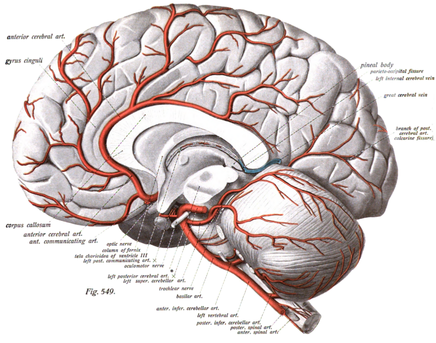
The anterior cerebral artery (ACA) is one of a pair of cerebral arteries that supplies oxygenated blood to most midline portions of the frontal lobes and superior medial parietal lobes of the brain. The two anterior cerebral arteries arise from the internal carotid artery and are part of the circle of Willis. The left and right anterior cerebral arteries are connected by the anterior communicating artery. Anterior cerebral artery syndrome refers to symptoms that follow a stroke occurring in the area normally supplied by one of the arteries. It is characterized by weakness and sensory loss in the lower leg and foot opposite to the lesion and behavioral changes. Structure The anterior cerebral artery is divided into 5 segments. Its smaller branches: the callosal (supracallosal) arteries are considered to be the A4 and A5 segments. *A1 originates from the internal carotid artery and extends to the ''anterior communicating artery'' (AComm). The ''anteromedial central'' (medial le ... [...More Info...] [...Related Items...] OR: [Wikipedia] [Google] [Baidu] |
Internal Capsule
The internal capsule is a white matter structure situated in the inferomedial part of each cerebral hemisphere of the brain. It carries information past the basal ganglia, separating the caudate nucleus and the thalamus from the putamen and the globus pallidus. The internal capsule contains both ascending and descending axons, going to and coming from the cerebral cortex. It also separates the caudate nucleus and the putamen in the dorsal striatum, a brain region involved in motor and reward pathways. The corticospinal tract constitutes a large part of the internal capsule, carrying motor information from the primary motor cortex to the lower motor neurons in the spinal cord. Above the basal ganglia the corticospinal tract is a part of the corona radiata. Below the basal ganglia the tract is called cerebral crus (a part of the cerebral peduncle) and below the pons it is referred to as the corticospinal tract. Structure The internal capsule consists of three parts and is V-sh ... [...More Info...] [...Related Items...] OR: [Wikipedia] [Google] [Baidu] |
Gray Substance
Grey matter is a major component of the central nervous system, consisting of neuronal cell bodies, neuropil (dendrites and unmyelinated axons), glial cells (astrocytes and oligodendrocytes), synapses, and capillaries. Grey matter is distinguished from white matter in that it contains numerous cell bodies and relatively few myelinated axons, while white matter contains relatively few cell bodies and is composed chiefly of long-range myelinated axons. The colour difference arises mainly from the whiteness of myelin. In living tissue, grey matter actually has a very light grey colour with yellowish or pinkish hues, which come from capillary blood vessels and neuronal cell bodies. Structure Grey matter refers to unmyelinated neurons and other cells of the central nervous system. It is present in the brain, brainstem and cerebellum, and present throughout the spinal cord. Grey matter is distributed at the surface of the cerebral hemispheres (cerebral cortex) and of the cerebel ... [...More Info...] [...Related Items...] OR: [Wikipedia] [Google] [Baidu] |


