|
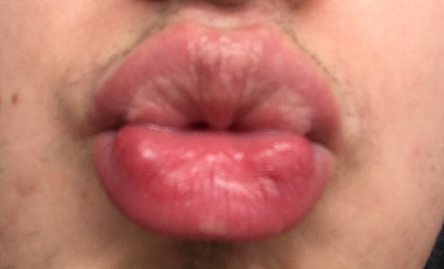
Angiokeratoma Of Mibelli
Angiokeratoma is a benign cutaneous lesion of capillaries, resulting in small marks of red to blue color and characterized by hyperkeratosis. ''Angiokeratoma corporis diffusum'' refers to Fabry's disease, but this is usually considered a distinct condition. Signs and symptoms Presentation includes telangiectasia, acanthosis, and hyperkeratosis. Presentation can be solitary or systemic. Multiple angiokeratomas, especially on the trunk in young people, are typical for Fabry disease, genetic disorder connected with systemic complications. Complications In some instances nodular angiokeratomas can produce necrotic tissue and valleys that can harbor fungal, bacterial and viral infections. Infections can include staphylococcus. If the lesion becomes painful, begins draining fluids or pus, or begins to smell, consult a physician. In these instance a doctor may recommend excision and grafting. Pathophysiology Histology Angiokeratomas characteristically have large dilated blood vess ... [...More Info...] [...Related Items...] OR: [Wikipedia] [Google] [Baidu] |
Cutaneous
Skin is the layer of usually soft, flexible outer tissue covering the body of a vertebrate animal, with three main functions: protection, regulation, and sensation. Other animal coverings, such as the arthropod exoskeleton, have different developmental origin, structure and chemical composition. The adjective cutaneous means "of the skin" (from Latin ''cutis'' 'skin'). In mammals, the skin is an organ of the integumentary system made up of multiple layers of ectodermal tissue and guards the underlying muscles, bones, ligaments, and internal organs. Skin of a different nature exists in amphibians, reptiles, and birds. Skin (including cutaneous and subcutaneous tissues) plays crucial roles in formation, structure, and function of extraskeletal apparatus such as horns of bovids (e.g., cattle) and rhinos, cervids' antlers, giraffids' ossicones, armadillos' osteoderm, and os penis/os clitoris. All mammals have some hair on their skin, even marine mammals like whales, dolphins, a ... [...More Info...] [...Related Items...] OR: [Wikipedia] [Google] [Baidu] |
Dermatologist
Dermatology is the branch of medicine dealing with the skin.''Random House Webster's Unabridged Dictionary.'' Random House, Inc. 2001. Page 537. . It is a speciality with both medical and surgical aspects. A dermatologist is a specialist medical doctor who manages diseases related to skin, hair, nails, and some cosmetic problems. Etymology Attested in English in 1819, the word "dermatology" derives from the Greek δέρματος (''dermatos''), genitive of δέρμα (''derma''), "skin" (itself from δέρω ''dero'', "to flay") and -λογία '' -logia''. Neo-Latin ''dermatologia'' was coined in 1630, an anatomical term with various French and German uses attested from the 1730s. History In 1708, the first great school of dermatology became a reality at the famous Hôpital Saint-Louis in Paris, and the first textbooks (Willan's, 1798–1808) and atlases ( Alibert's, 1806–1816) appeared in print around the same time.Freedberg, et al. (2003). ''Fitzpatrick's Dermatology in ... [...More Info...] [...Related Items...] OR: [Wikipedia] [Google] [Baidu] |
List Of Cutaneous Conditions
Many skin conditions affect the human integumentary system—the organ system covering the entire surface of the body and composed of skin, hair, nails, and related muscle and glands. The major function of this system is as a barrier against the external environment. The skin weighs an average of four kilograms, covers an area of two square metres, and is made of three distinct layers: the epidermis, dermis, and subcutaneous tissue. The two main types of human skin are: glabrous skin, the hairless skin on the palms and soles (also referred to as the "palmoplantar" surfaces), and hair-bearing skin.Burns, Tony; ''et al''. (2006) ''Rook's Textbook of Dermatology CD-ROM''. Wiley-Blackwell. . Within the latter type, the hairs occur in structures called pilosebaceous units, each with hair follicle, sebaceous gland, and associated arrector pili muscle. In the embryo, the epidermis, hair, and glands form from the ectoderm, which is chemically influenced by the underlying mesoderm th ... [...More Info...] [...Related Items...] OR: [Wikipedia] [Google] [Baidu] |
Vascular Malformation
A vascular malformation is a blood vessel or lymph vessel abnormality. Vascular malformations are one of the classifications of vascular anomalies, the other grouping is vascular tumors. They may cause aesthetic problems as they have a growth cycle, and can continue to grow throughout life. Vascular malformations of the brain (VMBs) include those involving capillaries, and those involving the veins and arteries. Capillary malformations in the brain are known as cerebral cavernous malformations or ''capillary cavernous malformations'' (CCMs). Those involving the mix of vessels are known as cerebral arteriovenous malformations (AVMs or cAVMs). The arteriovenous type is the most common in the brain. Types A simple division of the vascular malformations is made into ''low-flow'' and ''high-flow'' types. Low-flow malformations involve a single type of blood or lymph vessel, and are known as ''simple vascular malformations''; high-flow malformations involve an artery. There are also ma ... [...More Info...] [...Related Items...] OR: [Wikipedia] [Google] [Baidu] |
Vein
Veins are blood vessels in humans and most other animals that carry blood towards the heart. Most veins carry deoxygenated blood from the tissues back to the heart; exceptions are the pulmonary and umbilical veins, both of which carry oxygenated blood to the heart. In contrast to veins, arteries carry blood away from the heart. Veins are less muscular than arteries and are often closer to the skin. There are valves (called ''pocket valves'') in most veins to prevent backflow. Structure Veins are present throughout the body as tubes that carry blood back to the heart. Veins are classified in a number of ways, including superficial vs. deep, pulmonary vs. systemic, and large vs. small. * Superficial veins are those closer to the surface of the body, and have no corresponding arteries. *Deep veins are deeper in the body and have corresponding arteries. *Perforator veins drain from the superficial to the deep veins. These are usually referred to in the lower limbs and feet. *Communic ... [...More Info...] [...Related Items...] OR: [Wikipedia] [Google] [Baidu] |
Dermis
The dermis or corium is a layer of skin between the epidermis (with which it makes up the cutis) and subcutaneous tissues, that primarily consists of dense irregular connective tissue and cushions the body from stress and strain. It is divided into two layers, the superficial area adjacent to the epidermis called the papillary region and a deep thicker area known as the reticular dermis.James, William; Berger, Timothy; Elston, Dirk (2005). ''Andrews' Diseases of the Skin: Clinical Dermatology'' (10th ed.). Saunders. Pages 1, 11–12. . The dermis is tightly connected to the epidermis through a basement membrane. Structural components of the dermis are collagen, elastic fibers, and extrafibrillar matrix.Marks, James G; Miller, Jeffery (2006). ''Lookingbill and Marks' Principles of Dermatology'' (4th ed.). Elsevier Inc. Page 8–9. . It also contains mechanoreceptors that provide the sense of touch and thermoreceptors that provide the sense of heat. In addition, hair follicles, ... [...More Info...] [...Related Items...] OR: [Wikipedia] [Google] [Baidu] |
Fordyce's Spots
Fordyce spots (also termed Fordyce granules)James, William; Berger, Timothy; Elston, Dirk (2005). ''Andrews' Diseases of the Skin: Clinical Dermatology''. (10th ed.). Saunders. . are visible sebaceous glands that are present in most individuals. They appear on the genitals and/or on the face and in the mouth. They appear as small, painless, raised, pale, red or white spots or bumps 1 to 3 mm in diameter that may appear on the scrotum, shaft of the penis or on the labia, as well as the inner surface (retromolar mucosa) and vermilion border of the lips of the face. They are not associated with any disease or illness, nor are they infectious but rather they represent a natural occurrence on the body. No treatment is therefore required. Persons with this condition sometimes consult a dermatologist because they are worried they may have a sexually transmitted disease (especially genital warts) or some form of cancer. [...More Info...] [...Related Items...] OR: [Wikipedia] [Google] [Baidu] |
John Addison Fordyce
John Addison Fordyce (born 16 February 1858 in Guernsey County, Ohio, died on 4 June 1925 in New York City) was an American dermatologist, whose name is associated with Fordyce's spot (also known as Fordyce's disease or Fordyce's lesion), Angiokeratoma of Fordyce, Brooke–Fordyce trichoepithelioma, and Fox–Fordyce disease. Fordyce graduated in 1881 with a degree in medicine from the Chicago Medical College. He began his career in Hot Springs, Arkansas, but would travel to Europe in 1886. There he studied dermatology in Vienna and Paris. He returned to the States and settled down in New York, where he was a specialist in dermatology and syphilis. From 1889 to 1893 he taught at the New York Polyclinic, and later he served as a professor at the Bellevue Hospital College and the Columbia University College of Physicians and Surgeons Columbia University Vagelos College of Physicians and Surgeons (VP&S) is the graduate medical school of Columbia University, located at the Columbi ... [...More Info...] [...Related Items...] OR: [Wikipedia] [Google] [Baidu] |
Who Named It
''Whonamedit?'' is an online English-language dictionary of medical eponyms and the people associated with their identification. Though it is a dictionary, many eponyms and persons are presented in extensive articles with comprehensive bibliographies. The dictionary is hosted in Norway and maintained by medical historian Ole Daniel Enersen Ole Daniel Enersen (born March 14, 1943, in Oslo, Norway) is a Norwegian climber, photographer, journalist, writer, and medical historian. In 1965 he made the first ascent of the Trollveggen mountain in Romsdalen, Norway, along with Leif Norman .... References External links * Medical websites Medical dictionaries Eponyms {{online-dict-stub ... [...More Info...] [...Related Items...] OR: [Wikipedia] [Google] [Baidu] |
Vittorio Mibelli
Vittorio Mibelli (18 February 1860 – 26 April 1910) was an Italian dermatologist born in Portoferraio, Elba. He studied in Siena and Florence, afterwards working in Siena as a prosector at the anatomical institute and later as an assistant at the dermatology clinic. In 1888 he gained his habilitation, spending the following year in Hamburg, working with dermatologist Paul Gerson Unna (1850–1929). In 1890 he became an associate professor and director of dermatology clinic at the University of Cagliari. Two years later, he relocated to Parma, where he held the title of full professor from 1900 until his death in 1910 . Works His name is associated with two skin disorders: angiokeratoma of Mibelli and porokeratosis of Mibelli. Mibelli's disease I @ |
Biopsy
A biopsy is a medical test commonly performed by a surgeon, interventional radiologist, or an interventional cardiologist. The process involves extraction of sample cells or tissues for examination to determine the presence or extent of a disease. The tissue is then fixed, dehydrated, embedded, sectioned, stained and mounted before it is generally examined under a microscope by a pathologist; it may also be analyzed chemically. When an entire lump or suspicious area is removed, the procedure is called an excisional biopsy. An incisional biopsy or core biopsy samples a portion of the abnormal tissue without attempting to remove the entire lesion or tumor. When a sample of tissue or fluid is removed with a needle in such a way that cells are removed without preserving the histological architecture of the tissue cells, the procedure is called a needle aspiration biopsy. Biopsies are most commonly performed for insight into possible cancerous or inflammatory conditions. History T ... [...More Info...] [...Related Items...] OR: [Wikipedia] [Google] [Baidu] |
Hyperkeratosis
Hyperkeratosis is thickening of the stratum corneum (the outermost layer of the epidermis, or skin), often associated with the presence of an abnormal quantity of keratin,Kumar, Vinay; Fausto, Nelso; Abbas, Abul (2004) ''Robbins & Cotran Pathologic Basis of Disease'' (7th ed.). Saunders. Page 1230. . and also usually accompanied by an increase in the granular layer. As the corneum layer normally varies greatly in thickness in different sites, some experience is needed to assess minor degrees of hyperkeratosis. It can be caused by vitamin A deficiency or chronic exposure to arsenic. Hyperkeratosis can also be caused by B-Raf inhibitor drugs such as Vemurafenib and Dabrafenib.Niezgoda, Anna; Niezgoda, Piotr; Czajkowski, Rafal (2015) ''Novel Approaches to Treatment of Advanced Melanoma: A Review of Targeted Therapy and Immunotherapy'' BioMed Research International It can be treated with urea-containing creams, which dissolve the intercellular matrix of the cells of the stratum co ... [...More Info...] [...Related Items...] OR: [Wikipedia] [Google] [Baidu] |


