|
Alveolar Lung Disease
Alveolar lung diseases, are a group of diseases that mainly affect the alveoli of the lungs. Causes Alveoli are the functional units of the lungs. Alveolar lung diseases are classified as processes that affect these units that ultimately lead to issues with ventilation. There are a number of different causes of insult to the alveoli including build up of fluid, hemorrhage, infection, malignancy and build up of protein and mineral deposits. Conditions classified under alveolar lung disease include pulmonary edema (cardiogenic or non-cardiogenic), pneumonia (bacterial or viral), bronchoalveolar carcinoma, pulmonary hemorrhage, alveolar proteinosis and amyloidosis, and alveolar microlithiasis. Alveolar lung disease may be divided into acute or chronic. Causes of acute alveolar lung disease include pulmonary edema (cardiogenic or neurogenic), pneumonia (bacterial or viral), systemic lupus erythematosus, bleeding in the lungs (e.g., Goodpasture syndrome), idiopathic pulmonary hemos ... [...More Info...] [...Related Items...] OR: [Wikipedia] [Google] [Baidu] |
Pulmonary Alveolus
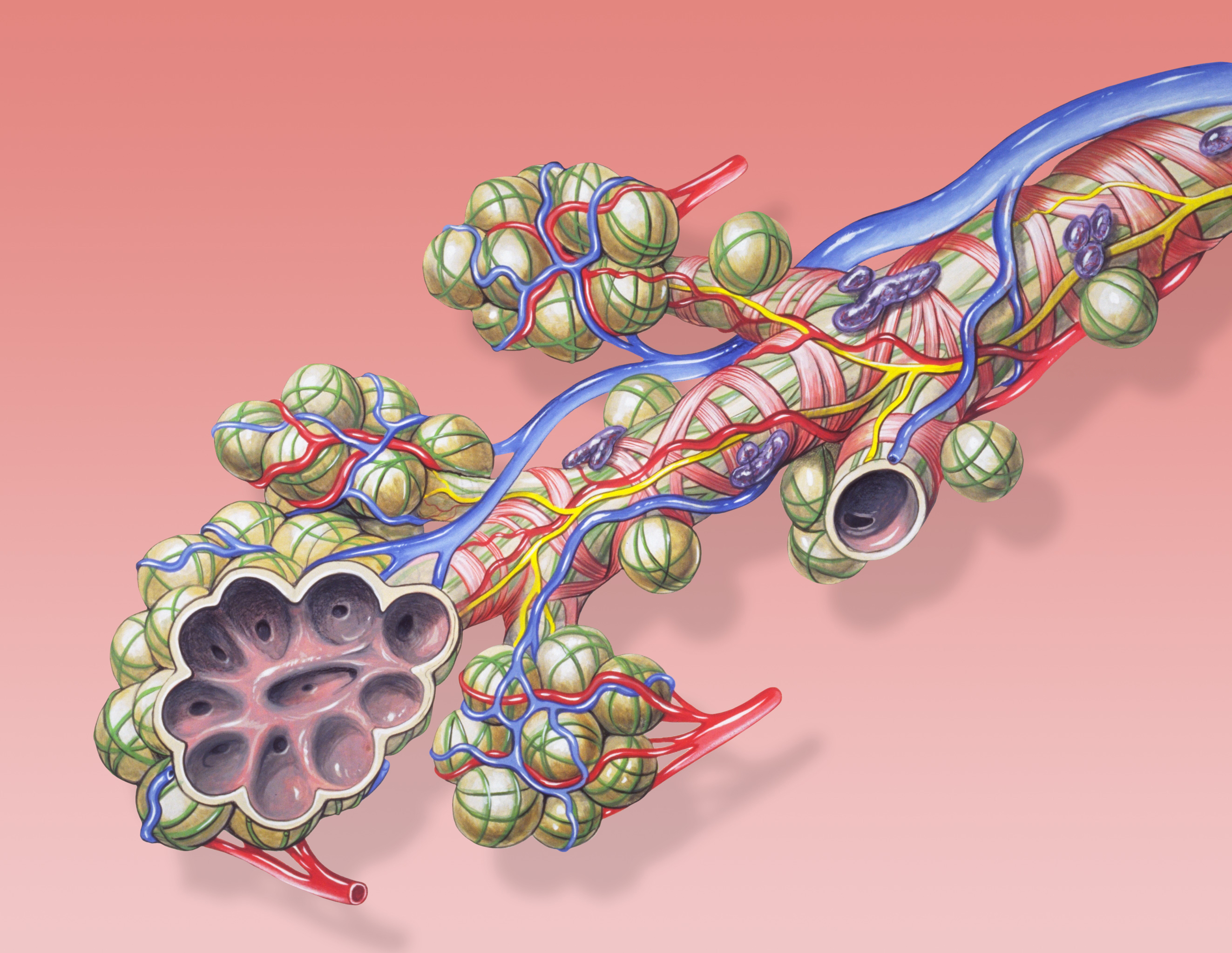
A pulmonary alveolus (plural: alveoli, from Latin ''alveolus'', "little cavity"), also known as an air sac or air space, is one of millions of hollow, distensible cup-shaped cavities in the lungs where oxygen is exchanged for carbon dioxide. Alveoli make up the functional tissue of the mammalian lungs known as the lung parenchyma, which takes up 90 percent of the total lung volume. Alveoli are first located in the respiratory bronchioles that mark the beginning of the respiratory zone. They are located sparsely in these bronchioles, line the walls of the alveolar ducts, and are more numerous in the blind-ended alveolar sacs. The acini are the basic units of respiration, with gas exchange taking place in all the alveoli present. The alveolar membrane is the gas exchange surface, surrounded by a network of capillaries. Across the membrane oxygen is diffused into the capillaries and carbon dioxide released from the capillaries into the alveoli to be breathed out. Alveoli are ... [...More Info...] [...Related Items...] OR: [Wikipedia] [Google] [Baidu] |
Non-invasive Ventilation
Non-invasive ventilation (NIV) is the use of breathing support administered through a face mask, nasal mask, or a helmet. Air, usually with added oxygen, is given through the mask under positive pressure; generally the amount of pressure is alternated depending on whether someone is breathing in or out. It is termed "non-invasive" because it is delivered with a mask that is tightly fitted to the face or around the head, but without a need for tracheal intubation (a tube through the mouth into the windpipe). While there are similarities with regard to the interface, NIV is not the same as continuous positive airway pressure (CPAP), which applies a single level of positive airway pressure throughout the whole respiratory cycle; CPAP does not deliver ventilation but is occasionally used in conditions also treated with NIV. Non-invasive ventilation is used in acute respiratory failure caused by a number of medical conditions, most prominently chronic obstructive pulmonary disease (CO ... [...More Info...] [...Related Items...] OR: [Wikipedia] [Google] [Baidu] |
Acute Respiratory Distress Syndrome
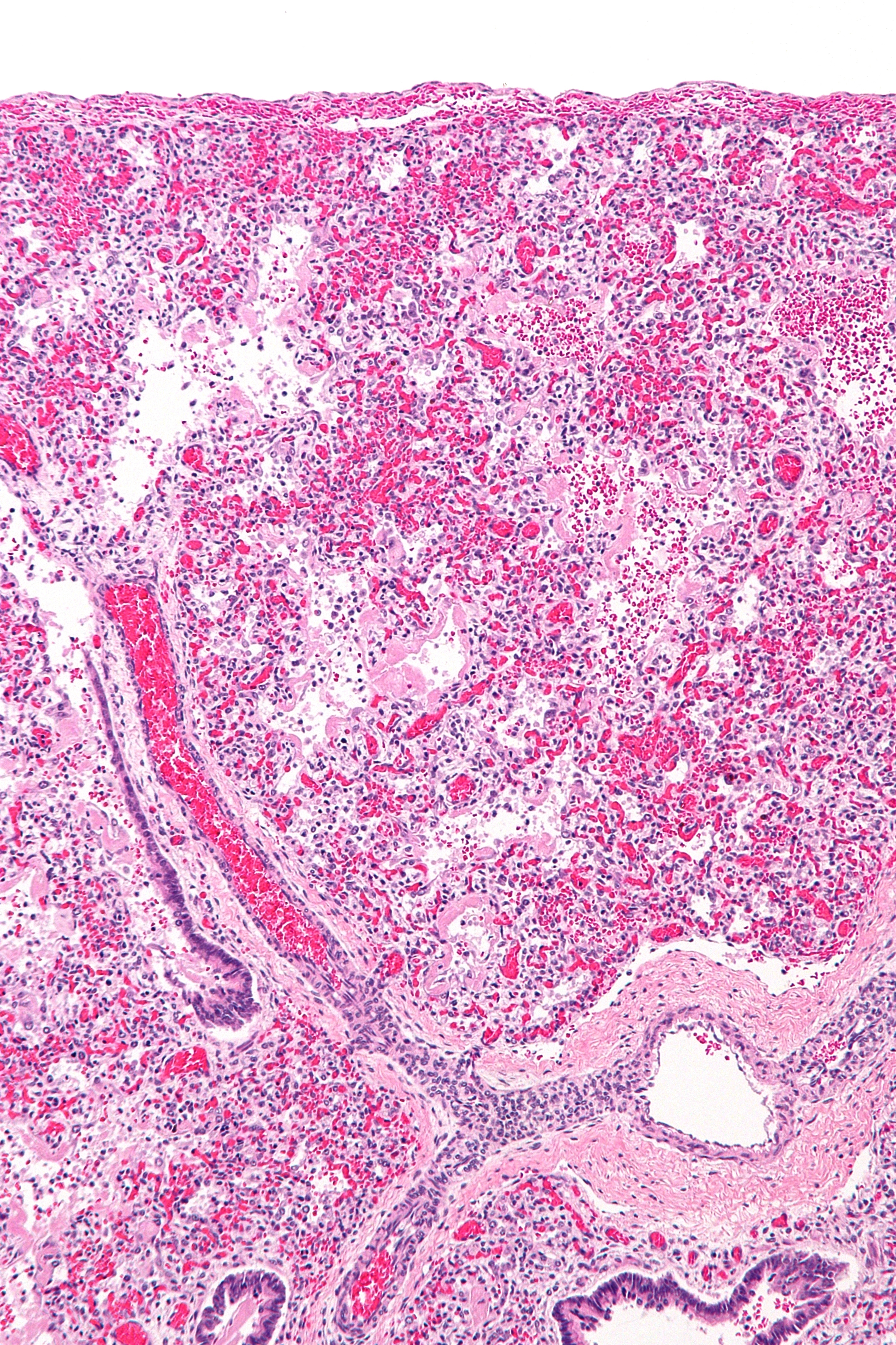
Acute respiratory distress syndrome (ARDS) is a type of respiratory failure characterized by rapid onset of widespread inflammation in the lungs. Symptoms include shortness of breath (dyspnea), rapid breathing (tachypnea), and bluish skin coloration (cyanosis). For those who survive, a decreased quality of life is common. Causes may include sepsis, pancreatitis, trauma, pneumonia, and aspiration. The underlying mechanism involves diffuse injury to cells which form the barrier of the microscopic air sacs of the lungs, surfactant dysfunction, activation of the immune system, and dysfunction of the body's regulation of blood clotting. In effect, ARDS impairs the lungs' ability to exchange oxygen and carbon dioxide. Adult diagnosis is based on a PaO2/FiO2 ratio (ratio of partial pressure arterial oxygen and fraction of inspired oxygen) of less than 300 mm Hg despite a positive end-expiratory pressure (PEEP) of more than 5 cm H2O. Cardiogenic pulmonary edema, as t ... [...More Info...] [...Related Items...] OR: [Wikipedia] [Google] [Baidu] |
Infarction
Infarction is tissue death ( necrosis) due to inadequate blood supply to the affected area. It may be caused by artery blockages, rupture, mechanical compression, or vasoconstriction. The resulting lesion is referred to as an infarct (from the Latin ''infarctus'', "stuffed into"). Causes Infarction occurs as a result of prolonged ischemia, which is the insufficient supply of oxygen and nutrition to an area of tissue due to a disruption in blood supply. The blood vessel supplying the affected area of tissue may be blocked due to an obstruction in the vessel (e.g., an arterial embolus, thrombus, or atherosclerotic plaque), compressed by something outside of the vessel causing it to narrow (e.g., tumor, volvulus, or hernia), ruptured by trauma causing a loss of blood pressure downstream of the rupture, or vasoconstricted, which is the narrowing of the blood vessel by contraction of the muscle wall rather than an external force (e.g., cocaine vasoconstriction le ... [...More Info...] [...Related Items...] OR: [Wikipedia] [Google] [Baidu] |
Pulmonary Embolism
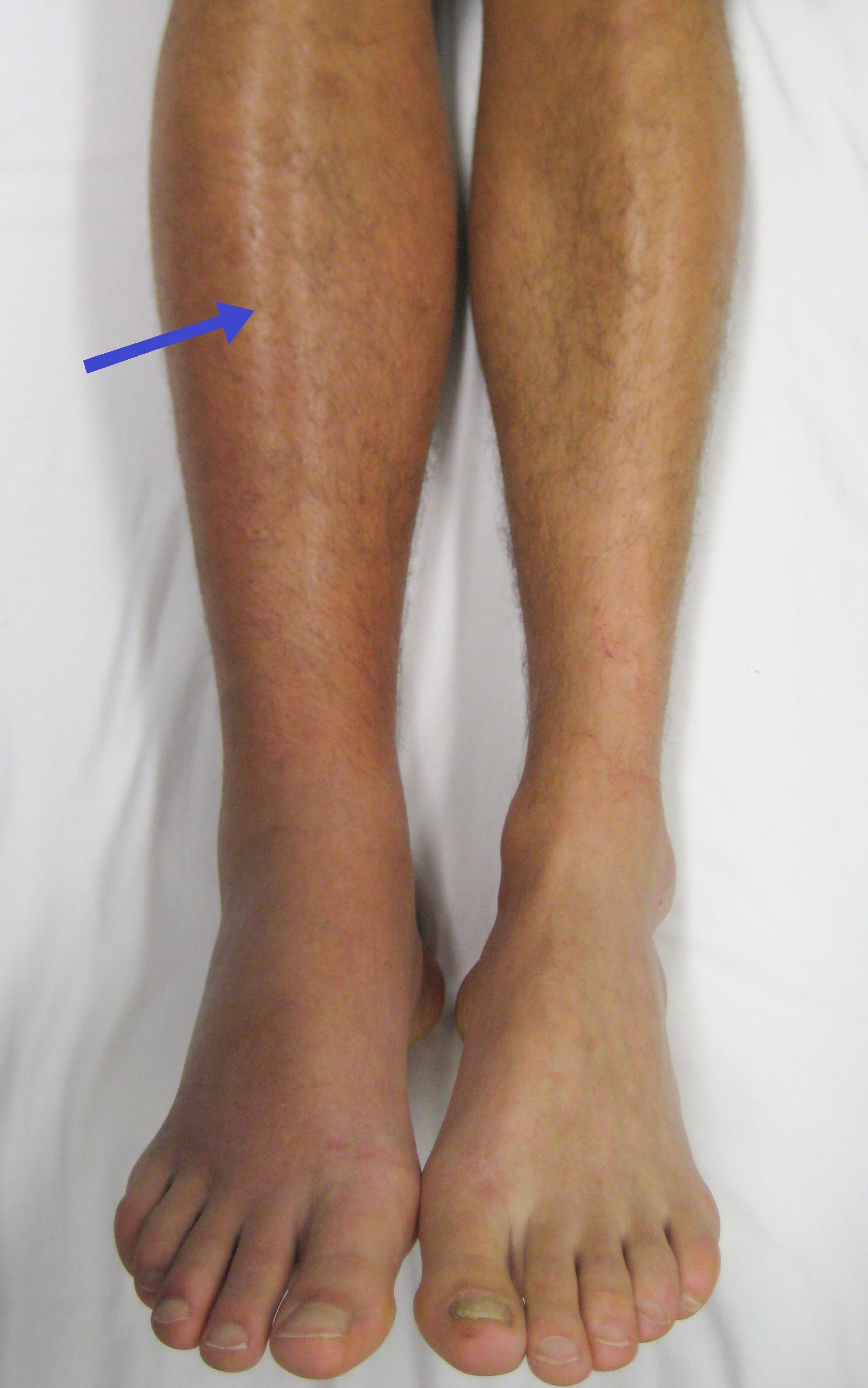
Pulmonary embolism (PE) is a blockage of an artery in the lungs by a substance that has moved from elsewhere in the body through the bloodstream ( embolism). Symptoms of a PE may include shortness of breath, chest pain particularly upon breathing in, and coughing up blood. Symptoms of a blood clot in the leg may also be present, such as a red, warm, swollen, and painful leg. Signs of a PE include low blood oxygen levels, rapid breathing, rapid heart rate, and sometimes a mild fever. Severe cases can lead to passing out, abnormally low blood pressure, obstructive shock, and sudden death. PE usually results from a blood clot in the leg that travels to the lung. The risk of blood clots is increased by advanced age, cancer, prolonged bed rest and immobilization, smoking, stroke, long-haul travel over 4 hours, certain genetic conditions, estrogen-based medication, pregnancy, obesity, trauma or bone fracture, and after some types of surgery. A small proportion of cases are ... [...More Info...] [...Related Items...] OR: [Wikipedia] [Google] [Baidu] |
Lung Contusion
The lungs are the primary organs of the respiratory system in humans and most other animals, including some snails and a small number of fish. In mammals and most other vertebrates, two lungs are located near the backbone on either side of the heart. Their function in the respiratory system is to extract oxygen from the air and transfer it into the bloodstream, and to release carbon dioxide from the bloodstream into the atmosphere, in a process of gas exchange. Respiration is driven by different muscular systems in different species. Mammals, reptiles and birds use their different muscles to support and foster breathing. In earlier tetrapods, air was driven into the lungs by the pharyngeal muscles via buccal pumping, a mechanism still seen in amphibians. In humans, the main muscle of respiration that drives breathing is the diaphragm. The lungs also provide airflow that makes vocal sounds including human speech possible. Humans have two lungs, one on the left and on ... [...More Info...] [...Related Items...] OR: [Wikipedia] [Google] [Baidu] |
Atelectasis
Atelectasis is the collapse or closure of a lung resulting in reduced or absent gas exchange. It is usually unilateral, affecting part or all of one lung. It is a condition where the alveoli are deflated down to little or no volume, as distinct from pulmonary consolidation, in which they are filled with liquid. It is often called a ''collapsed lung'', although that term may also refer to pneumothorax. It is a very common finding in chest X-rays and other radiological studies, and may be caused by normal exhalation or by various medical conditions. Although frequently described as a ''collapse of lung tissue'', atelectasis is not synonymous with a pneumothorax, which is a more specific condition that can cause atelectasis. Acute atelectasis may occur as a post-operative complication or as a result of surfactant deficiency. In premature babies, this leads to infant respiratory distress syndrome. The term uses combining forms of ''atel-'' + '' ectasis'', from el, ἀτελ ... [...More Info...] [...Related Items...] OR: [Wikipedia] [Google] [Baidu] |
Aspiration Pneumonia
Aspiration pneumonia is a type of lung infection that is due to a relatively large amount of material from the stomach or mouth entering the lungs. Signs and symptoms often include fever and cough of relatively rapid onset. Complications may include lung abscess, acute respiratory distress syndrome, empyema, and parapneumonic effusion. Some include chemical induced inflammation of the lungs as a subtype, which occurs from acidic but non-infectious stomach contents entering the lungs. Infection can be due to a variety of bacteria. Risk factors include decreased level of consciousness, problems with swallowing, alcoholism, tube feeding, and poor oral health. Diagnosis is typically based on the presenting history, symptoms, chest X-ray, and sputum culture. Differentiating from other types of pneumonia may be difficult. Treatment is typically with antibiotics such as clindamycin, meropenem, ampicillin/sulbactam, or moxifloxacin. For those with only chemical pneumonitis, anti ... [...More Info...] [...Related Items...] OR: [Wikipedia] [Google] [Baidu] |
Air Bronchogram
An air bronchogram is defined as a pattern of air-filled bronchi on a background of airless lung. Material was copied from this source, which is available under Creative Commons Attribution 4.0 International License __TOC__ Consolidations In pulmonary consolidations and infiltrates, air bronchograms are most commonly caused by pneumonia or pulmonary edema (especially with alveolar edema). Other potential causes of consolidations or infiltrates with air bronchograms are: * Pulmonary edema * Non-obstructive atelectasis * Severe interstitial lung disease * Pulmonary infarct * Pulmonary hemorrhage * Normal expiration Lung nodules For lung nodules, air bronchograms used to be associated with infectious causes of consolidation and, therefore to be benign. However, in the setting of a lung nodule, an air bronchogram is actually more frequent in malignant than in benign nodules. studied the tumour-bronchus relationship and described five types: * In “Type 1” the bronchial lumen is ... [...More Info...] [...Related Items...] OR: [Wikipedia] [Google] [Baidu] |
Pulse Oximetry
Pulse oximetry is a noninvasive method for monitoring a person's oxygen saturation. Peripheral oxygen saturation (SpO2) readings are typically within 2% accuracy (within 4% accuracy in 95% of cases) of the more accurate (and invasive) reading of arterial oxygen saturation (SaO2) from arterial blood gas analysis. But the two are correlated well enough that the safe, convenient, noninvasive, inexpensive pulse oximetry method is valuable for measuring oxygen saturation in clinical use. The most common approach is ''transmissive pulse oximetry''. In this approach, a sensor device is placed on a thin part of the patient's body, usually a fingertip or earlobe, or an infant's foot. Fingertips and earlobes have higher blood flow rates than other tissues, which facilitates heat transfer. The device passes two wavelengths of light through the body part to a photodetector. It measures the changing absorbance at each of the wavelengths, allowing it to determine the absorbances due to t ... [...More Info...] [...Related Items...] OR: [Wikipedia] [Google] [Baidu] |
Metastases
Metastasis is a pathogenic agent's spread from an initial or primary site to a different or secondary site within the host's body; the term is typically used when referring to metastasis by a cancerous tumor. The newly pathological sites, then, are metastases (mets). It is generally distinguished from cancer invasion, which is the direct extension and penetration by cancer cells into neighboring tissues. Cancer occurs after cells are genetically altered to proliferate rapidly and indefinitely. This uncontrolled proliferation by mitosis produces a primary heterogeneic tumour. The cells which constitute the tumor eventually undergo metaplasia, followed by dysplasia then anaplasia, resulting in a malignant phenotype. This malignancy allows for invasion into the circulation, followed by invasion to a second site for tumorigenesis. Some cancer cells known as circulating tumor cells acquire the ability to penetrate the walls of lymphatic vessel, lymphatic or blood vessels, after ... [...More Info...] [...Related Items...] OR: [Wikipedia] [Google] [Baidu] |


