|
Agouti-signaling Protein
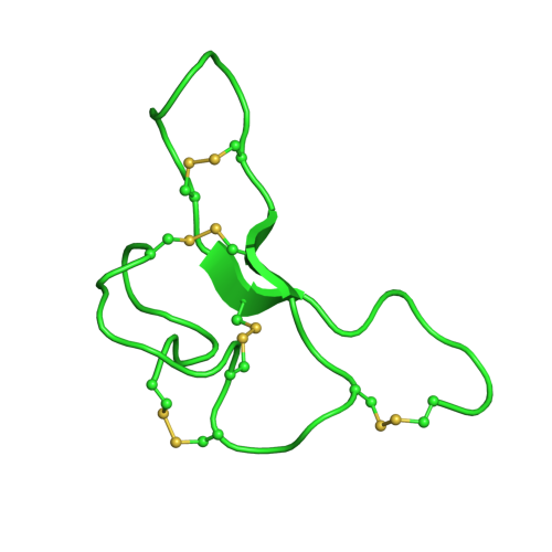
Agouti-signaling protein is a protein that in humans is encoded by the ASIP gene. It is responsible for the distribution of melanin pigment in mammals. Agouti interacts with the melanocortin 1 receptor to determine whether the melanocyte (pigment cell) produces phaeomelanin (a red to yellow pigment), or eumelanin (a brown to black pigment). This interaction is responsible for making distinct light and dark bands in the hairs of animals such as the agouti, which the gene is named after. In other species such as horses, agouti signalling is responsible for determining which parts of the body will be red or black. Mice with wildtype agouti will be grey, with each hair being partly yellow and partly black. Loss of function mutations in mice and other species cause black fur coloration, while mutations causing expression throughout the whole body in mice cause yellow fur and obesity. The agouti-signaling protein (ASIP) is a competitive antagonist with alpha-Melanocyte-stimulating hormon ... [...More Info...] [...Related Items...] OR: [Wikipedia] [Google] [Baidu] |
Protein
Proteins are large biomolecules and macromolecules that comprise one or more long chains of amino acid residues. Proteins perform a vast array of functions within organisms, including catalysing metabolic reactions, DNA replication, responding to stimuli, providing structure to cells and organisms, and transporting molecules from one location to another. Proteins differ from one another primarily in their sequence of amino acids, which is dictated by the nucleotide sequence of their genes, and which usually results in protein folding into a specific 3D structure that determines its activity. A linear chain of amino acid residues is called a polypeptide. A protein contains at least one long polypeptide. Short polypeptides, containing less than 20–30 residues, are rarely considered to be proteins and are commonly called peptides. The individual amino acid residues are bonded together by peptide bonds and adjacent amino acid residues. The sequence of amino acid resid ... [...More Info...] [...Related Items...] OR: [Wikipedia] [Google] [Baidu] |
Alpha-Melanocyte-stimulating Hormone
α-Melanocyte-stimulating hormone (α-MSH) is an endogenous peptide hormone and neuropeptide of the melanocortin family, with a tridecapeptide structure and the amino acid sequence Ac-Ser-Tyr-Ser-Met-Glu-His-Phe-Arg-Trp-Gly-Lys-Pro-Val-NH2. It is the most important of the melanocyte-stimulating hormones (MSHs) (also known as melanotropins) in stimulating melanogenesis, a process that in mammals (including humans) is responsible for pigmentation primarily of the hair and skin. It also plays a role in feeding behavior, energy homeostasis, sexual activity, and protection against ischemia and reperfusion injury. α-MSH is a non-selective full agonist of the melanocortin receptors MC1 (Ki = 0.230 nM), MC3 (Ki = 31.5 nM), MC4 (Ki = 900 nM), and MC5 (Ki = 7160 nM), but not MC2 (which is exclusive for adrenocorticotropic hormone (ACTH)). Activation of the MC1 receptor is responsible for its effect on pigmentation, whereas its regulation of appetite, metabolism, and sexual behavi ... [...More Info...] [...Related Items...] OR: [Wikipedia] [Google] [Baidu] |
Ectopic Expression
Ectopic is a word used with a prefix, ecto, meaning “out of place.” Ectopic expression is an abnormal gene expression in a cell type, tissue type, or developmental stage in which the gene is not usually expressed. The term ectopic expression is predominantly used in studies using metazoans, especially in ''Drosophila melanogaster'' for research purposes. How is it used Although ectopic expression can be caused by a natural condition, it is uncommonly seen in nature because it is a product of defects in gene regulation. In fact, ectopic expression is more commonly used for research purposes. Artificially induced gene expression helps to determine the function of a gene of interest. Common techniques such as overexpressing or misexpressing the genes by ''UA' system in ''D. melanogaster'' are used. In model organisms, such techniques are used to perform genetic screens to identify a function of the gene involved in specific cellular or developmental processes. Ectopic expression ... [...More Info...] [...Related Items...] OR: [Wikipedia] [Google] [Baidu] |
Tissue (biology)
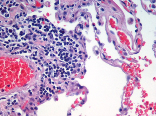
In biology, tissue is a biological organizational level between cells and a complete organ. A tissue is an ensemble of similar cells and their extracellular matrix from the same origin that together carry out a specific function. Organs are then formed by the functional grouping together of multiple tissues. The English word "tissue" derives from the French word "tissu", the past participle of the verb tisser, "to weave". The study of tissues is known as histology or, in connection with disease, as histopathology. Xavier Bichat is considered as the "Father of Histology". Plant histology is studied in both plant anatomy and physiology. The classical tools for studying tissues are the paraffin block in which tissue is embedded and then sectioned, the histological stain, and the optical microscope. Developments in electron microscopy, immunofluorescence, and the use of frozen tissue-sections have enhanced the detail that can be observed in tissues. With these tools, th ... [...More Info...] [...Related Items...] OR: [Wikipedia] [Google] [Baidu] |
Trophoblast
The trophoblast (from Greek : to feed; and : germinator) is the outer layer of cells of the blastocyst. Trophoblasts are present four days after fertilization in humans. They provide nutrients to the embryo and develop into a large part of the placenta. They form during the first stage of pregnancy and are the first cells to differentiate from the fertilized egg to become extraembryonic structures that do not directly contribute to the embryo. After gastrulation, the trophoblast is contiguous with the ectoderm of the embryo and is referred to as the trophectoderm. After the first differentiation, the cells in the human embryo lose their totipotency and are no longer totipotent stem cells because they cannot form a trophoblast. They are now pluripotent stem cells. Structure The trophoblast proliferates and differentiates into two cell layers at approximately six days after fertilization for humans. Function Trophoblasts are specialized cells of the placenta that play a ... [...More Info...] [...Related Items...] OR: [Wikipedia] [Google] [Baidu] |
Agouti-related Peptide
Agouti-related protein (AgRP), also called agouti-related peptide, is a neuropeptide produced in the brain by the AgRP/NPY neuron. It is synthesized in neuropeptide Y (NPY)-containing cell bodies located in the ventromedial part of the arcuate nucleus in the hypothalamus. AgRP is co-expressed with NPY and acts to increase appetite and decrease metabolism and energy expenditure. It is one of the most potent and long-lasting of appetite stimulators. In humans, the agouti-related peptide is encoded by the ''AGRP'' gene. Structure AgRP is a paracrine signaling molecule made of 112 amino acids (the gene product of 132 amino acids is processed by removal of the N-terminal 20-residue signal peptide domain). It was independently identified by two teams in 1997 based on its sequence similarity with agouti signalling peptide (ASIP), a protein synthesized in the skin controlling coat colour. AgRP is approximately 25% identical to ASIP. The murine homologue of AgRP consists of 111 amin ... [...More Info...] [...Related Items...] OR: [Wikipedia] [Google] [Baidu] |
Inhibitor Cystine Knot
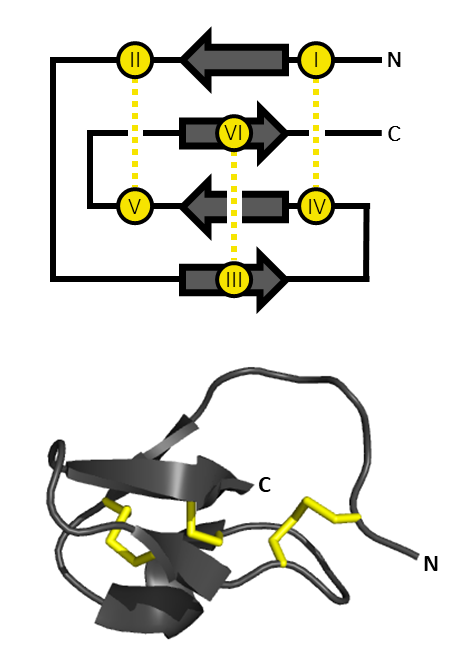
An inhibitor cystine knot (aka ICK or Knottin) is a protein structural motif containing three disulfide bridges. Knottins are one of three folds in the cystine knot motif; the other closely related knots are the Growth Factor Cystine Knot (GFCK) and the Cyclic Cystine Knot (CCK; cyclotide). Types include a) cyclic mobius, b) cyclic bracelet, c) acyclic inhibitor knottins. Cystine knot motifs are found frequently in nature in a plethora of plants, animals, and fungi and serve diverse functions from appetite suppression to anti-fungal activity. Along with the sections of polypeptide between them, two disulfides form a loop through which the third disulfide bond (linking the 3rd and 6th cysteine in the sequence) passes, forming a knot. The motif is common in invertebrate toxins such as those from arachnids and molluscs. The motif is also found in some inhibitor proteins found in plants, but the plant and animal motifs are thought to be a product of convergent evolution. The ICK moti ... [...More Info...] [...Related Items...] OR: [Wikipedia] [Google] [Baidu] |
Murinae
The Old World rats and mice, part of the subfamily Murinae in the family Muridae, comprise at least 519 species. Members of this subfamily are called murines. In terms of species richness, this subfamily is larger than all mammal families except the Cricetidae and Muridae, and is larger than all mammal orders except the bats and the remainder of the rodents. Description The Murinae are native to Africa, Europe, Asia, and Australia. They are terrestrial placental mammals. They have also been introduced to all continents except Antarctica, and are serious pest animals. This is particularly true in island communities where they have contributed to the endangerment and extinction of many native animals. Two prominent murine species have become vital laboratory animals: the brown rat and house mouse are both used as medical subjects. The murines have a distinctive molar pattern that involves three rows of cusps instead of two, the primitive pattern seen most fr ... [...More Info...] [...Related Items...] OR: [Wikipedia] [Google] [Baidu] |
Tumorigenesis
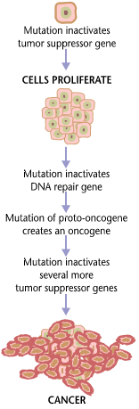
Carcinogenesis, also called oncogenesis or tumorigenesis, is the formation of a cancer, whereby normal cells are transformed into cancer cells. The process is characterized by changes at the cellular, genetic, and epigenetic levels and abnormal cell division. Cell division is a physiological process that occurs in almost all tissues and under a variety of circumstances. Normally, the balance between proliferation and programmed cell death, in the form of apoptosis, is maintained to ensure the integrity of tissues and organs. According to the prevailing accepted theory of carcinogenesis, the somatic mutation theory, mutations in DNA and epimutations that lead to cancer disrupt these orderly processes by interfering with the programming regulating the processes, upsetting the normal balance between proliferation and cell death. This results in uncontrolled cell division and the evolution of those cells by natural selection in the body. Only certain mutations lead to can ... [...More Info...] [...Related Items...] OR: [Wikipedia] [Google] [Baidu] |
Hyperinsulinemia
Hyperinsulinemia is a condition in which there are excess levels of insulin circulating in the blood relative to the level of glucose. While it is often mistaken for diabetes or hyperglycaemia, hyperinsulinemia can result from a variety of metabolic diseases and conditions, as well as non-nutritive sugars in the diet. While hyperinsulinemia is often seen in people with early stage type 2 diabetes mellitus, it is not the cause of the condition and is only one symptom of the disease. Type 1 diabetes only occurs when pancreatic beta-cell function is impaired. Hyperinsulinemia can be seen in a variety of conditions including diabetes mellitus type 2, in neonates and in drug-induced hyperinsulinemia. It can also occur in congenital hyperinsulinism, including nesidioblastosis. Hyperinsulinemia is associated with hypertension, obesity, dyslipidemia, insulin resistance, and glucose intolerance. These conditions are collectively known as metabolic syndrome. This close association between h ... [...More Info...] [...Related Items...] OR: [Wikipedia] [Google] [Baidu] |
Wild Type
The wild type (WT) is the phenotype of the typical form of a species as it occurs in nature. Originally, the wild type was conceptualized as a product of the standard "normal" allele at a locus, in contrast to that produced by a non-standard, "mutant" allele. "Mutant" alleles can vary to a great extent, and even become the wild type if a genetic shift occurs within the population. Continued advancements in genetic mapping technologies have created a better understanding of how mutations occur and interact with other genes to alter phenotype. It is now appreciated that most or all gene loci exist in a variety of allelic forms, which vary in frequency throughout the geographic range of a species, and that a uniform wild type does not exist. In general, however, the most prevalent allele – i.e., the one with the highest gene frequency – is the one deemed wild type. The concept of wild type is useful in some experimental organisms such as fruit flies ''Drosophila melanogaster'' ... [...More Info...] [...Related Items...] OR: [Wikipedia] [Google] [Baidu] |


