|
Accessory Meningeal Artery
The accessory meningeal artery (also accessory branch of middle meningeal artery, pterygomeningeal artery, small meningeal or parvidural branch) is a branch of the maxillary artery, sometimes derived from the middle meningeal artery. Course It enters the skull through the Foramen ovale (skull), foramen ovale, and supplies the trigeminal ganglion and dura mater. Nomenclature Only about 10% of the blood flowing through this artery reaches intracranial structures. The remaining blood flow is dispersed to extracranial structures around the infratemporal fossa. Reflecting this fact, Terminologia Anatomica lists entries for both "accessory branch of middle meningeal artery" and "pterygomeningeal artery". References External links * * () Arteries of the head and neck {{circulatory-stub ... [...More Info...] [...Related Items...] OR: [Wikipedia] [Google] [Baidu] |
Maxillary Artery
The maxillary artery supplies deep structures of the face. It branches from the external carotid artery just deep to the neck of the mandible. Structure The maxillary artery, the larger of the two terminal branches of the external carotid artery, arises behind the neck of the mandible, and is at first imbedded in the substance of the parotid gland; it passes forward between the ramus of the mandible and the sphenomandibular ligament, and then runs, either superficial or deep to the lateral pterygoid muscle, to the pterygopalatine fossa. It supplies the deep structures of the face, and may be divided into mandibular, pterygoid, and pterygopalatine portions. First portion The ''first'' or ''mandibular '' or ''bony'' portion passes horizontally forward, between the neck of the mandible and the sphenomandibular ligament, where it lies parallel to and a little below the auriculotemporal nerve; it crosses the inferior alveolar nerve, and runs along the lower border of the lateral pte ... [...More Info...] [...Related Items...] OR: [Wikipedia] [Google] [Baidu] |
Meninges
In anatomy, the meninges (, ''singular:'' meninx ( or ), ) are the three membranes that envelop the brain and spinal cord. In mammals, the meninges are the dura mater, the arachnoid mater, and the pia mater. Cerebrospinal fluid is located in the subarachnoid space between the arachnoid mater and the pia mater. The primary function of the meninges is to protect the central nervous system. Structure Dura mater The dura mater ( la, tough mother) (also rarely called ''meninx fibrosa'' or ''pachymeninx'') is a thick, durable membrane, closest to the Human skull, skull and vertebrae. The dura mater, the outermost part, is a loosely arranged, fibroelastic layer of cells, characterized by multiple interdigitating cell processes, no extracellular collagen, and significant extracellular spaces. The middle region is a mostly fibrous portion. It consists of two layers: the endosteal layer, which lies closest to the Calvaria (skull), skull, and the inner meningeal layer, which lies closer ... [...More Info...] [...Related Items...] OR: [Wikipedia] [Google] [Baidu] |
Middle Meningeal Artery
The middle meningeal artery ('' la, arteria meningea media'') is typically the third branch of the first portion of the maxillary artery. After branching off the maxillary artery in the infratemporal fossa, it runs through the foramen spinosum to supply the dura mater (the outer meningeal layer) and the calvaria. The middle meningeal artery is the largest of the three (paired) arteries that supply the meninges, the others being the anterior meningeal artery and the posterior meningeal artery. The anterior branch of the middle meningeal artery runs beneath the pterion. It is vulnerable to injury at this point, where the skull is thin. Rupture of the artery may give rise to an epidural hematoma. In the dry cranium, the middle meningeal, which runs within the dura mater surrounding the brain, makes a deep groove in the calvarium. The middle meningeal artery is intimately associated with the auriculotemporal nerve, which wraps around the artery making the two easily identifiable i ... [...More Info...] [...Related Items...] OR: [Wikipedia] [Google] [Baidu] |
Foramen Ovale (skull)
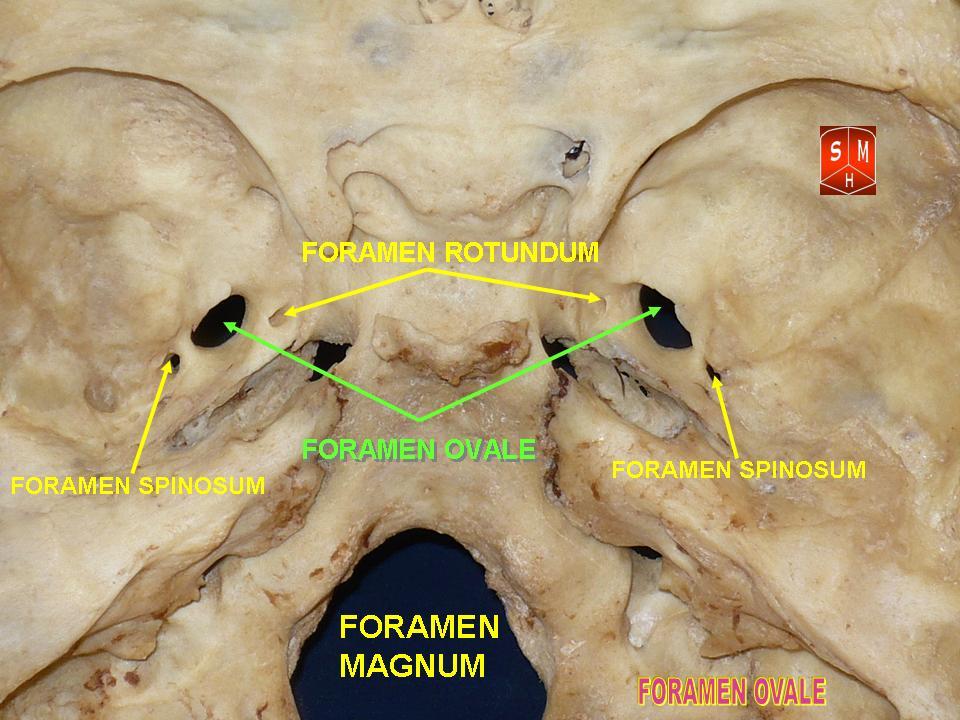
The foramen ovale (Latin: oval window) is a hole in the posterior part of the sphenoid bone, posterolateral to the foramen rotundum. It is one of the larger of the several holes (the foramina) in the skull. It transmits the mandibular nerve, a branch of the trigeminal nerve. Structure The foramen ovale is an opening in the greater wing of the sphenoid bone. The foramen ovale is one of two cranial foramina in the greater wing, the other being the foramen spinosum. The foramen ovale is posterolateral to the foramen rotundum and anteromedial to the foramen spinosum. Posterior and medial to the foramen is the opening for the carotid canal. Variation Similar to other foramina, the foramen ovale differs in shape and size throughout the natural life. The earliest perfect ring-shaped formation of the foramen ovale was observed in the 7th fetal month and the latest in 3 years after birth, in a study using over 350 skulls.In a study conducted on 100 skulls, the foramen ovale was d ... [...More Info...] [...Related Items...] OR: [Wikipedia] [Google] [Baidu] |
Trigeminal Ganglion
A trigeminal ganglion (or Gasserian ganglion, or semilunar ganglion, or Gasser's ganglion) is the sensory ganglion at the base of each of the two trigeminal nerves (CN V), occupying a cavity (Meckel's cave) in the dura mater, covering the trigeminal impression near the apex of the petrous part of the temporal bone. Structure It is somewhat crescent-shaped, with its convexity directed forward: Medially, it is in relation with the internal carotid artery and the posterior part of the cavernous sinus. The motor root runs in front of and medial to the sensory root, and passes beneath the ganglion; it leaves the skull through the foramen ovale, and, immediately below this foramen, joins the mandibular nerve. The greater superficial petrosal nerve lies also underneath the ganglion. The ganglion receives, on its medial side, filaments from the carotid plexus of the sympathetic. It gives off minute branches to the tentorium cerebelli, and to the dura mater in the middle fossa of th ... [...More Info...] [...Related Items...] OR: [Wikipedia] [Google] [Baidu] |
Dura Mater
In neuroanatomy, dura mater is a thick membrane made of dense irregular connective tissue that surrounds the brain and spinal cord. It is the outermost of the three layers of membrane called the meninges that protect the central nervous system. The other two meningeal layers are the arachnoid mater and the pia mater. It envelops the arachnoid mater, which is responsible for keeping in the cerebrospinal fluid. It is derived primarily from the neural crest cell population, with postnatal contributions of the paraxial mesoderm. Structure The dura mater has several functions and layers. The dura mater is a membrane that envelops the arachnoid mater. It surrounds and supports the dural sinuses (also called dural venous sinuses, cerebral sinuses, or cranial sinuses) and carries blood from the brain toward the heart. Cranial dura mater has two layers called ''lamellae'', a superficial layer (also called the periosteal layer), which serves as the skull's inner periosteum, called the ... [...More Info...] [...Related Items...] OR: [Wikipedia] [Google] [Baidu] |
Terminologia Anatomica
''Terminologia Anatomica'' is the international standard for human anatomical terminology. It is developed by the Federative International Programme on Anatomical Terminology, a program of the International Federation of Associations of Anatomists (IFAA). The second edition was released in 2019 and approved and adopted by the IFAA General Assembly in 2020. ''Terminologia Anatomica'' supersedes the previous standard, ''Nomina Anatomica''. It contains terminology for about 7500 human anatomical structures. Categories of anatomical structures ''Terminologia Anatomica'' is divided into 16 chapters grouped into five parts. The official terms are in Latin. Although equivalent English-language terms are provided, as shown below, only the official Latin terms are used as the basis for creating lists of equivalent terms in other languages. Part I Chapter 1: General anatomy # General terms # Reference planes # Reference lines # Human body positions # Movements # Parts of human body ... [...More Info...] [...Related Items...] OR: [Wikipedia] [Google] [Baidu] |


