|
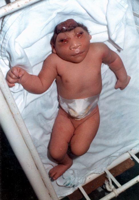
Acalvaria
Acalvaria is a rare malformation consisting of absence of the calvarial bones, dura mater and associated muscles in the presence of a normal skull base and normal facial bones. The central nervous system is usually unaffected. The presumed pathogenesis of acalvaria is faulty migration of the membranous neurocranium with normal placement of the embryonic ectoderm, resulting in absence of the calvaria but an intact layer of skin over the brain parenchyma. In other words, instead of having a skull cap protecting the brain, there is only skin covering it."Acalvaria." Right Diagnosis. Health Grades Inc., n.d. Web. 27 Nov. 2012. < ...
|
Anencephaly
Anencephaly is the absence of a major portion of the brain, skull, and scalp that occurs during embryonic development. It is a cephalic disorder that results from a neural tube defect that occurs when the rostral (head) end of the neural tube fails to close, usually between the 23rd and 26th day following conception. Strictly speaking, the Greek term translates as "without a brain" (or totally lacking the inside part of the head), but it is accepted that children born with this disorder usually only lack a telencephalon, the largest part of the brain consisting mainly of the cerebral hemispheres, including the neocortex, which is responsible for cognition. The remaining structure is usually covered only by a thin layer of membrane—skin, bone, meninges, etc., are all lacking. With very few exceptions, infants with this disorder do not survive longer than a few hours or days after birth. Signs and symptoms The National Institute of Neurological Disorders and Stroke (NINDS) descr ... [...More Info...] [...Related Items...] OR: [Wikipedia] [Google] [Baidu] |
Medical Genetics
Medical genetics is the branch tics in that human genetics is a field of scientific research that may or may not apply to medicine, while medical genetics refers to the application of genetics to medical care. For example, research on the causes and inheritance of genetic disorders would be considered within both human genetics and medical genetics, while the diagnosis, management, and counselling people with genetic disorders would be considered part of medical genetics. In contrast, the study of typically non-medical phenotypes such as the genetics of eye color would be considered part of human genetics, but not necessarily relevant to medical genetics (except in situations such as albinism). ''Genetic medicine'' is a newer term for medical genetics and incorporates areas such as gene therapy, personalized medicine, and the rapidly emerging new medical specialty, predictive medicine. Scope Medical genetics encompasses many different areas, including clinical practice of ... [...More Info...] [...Related Items...] OR: [Wikipedia] [Google] [Baidu] |
Calvaria (skull)
The calvaria is the top part of the skull. It is the upper part of the neurocranium and covers the cranial cavity containing the brain. It forms the main component of the skull roof. The calvaria is made up of the superior portions of the frontal bone, occipital bone, and parietal bones. In the human skull, the sutures between the bones normally remain flexible during the first few years of postnatal development, and fontanelles are palpable. Premature complete ossification of these sutures is called craniosynostosis. In Latin, the word ''calvaria'' is used as a feminine noun with plural ''calvariae''; however, many medical texts list the word as ''calvarium'', a neuter Latin noun with plural ''calvaria''. Structure The outer surface of the skull possesses a number of landmarks. The point at which the frontal bone and the two parietal bones meet is known as "Bregma". The point at which the two parietal bones and the occipital bone meet is known as "Lambda". Not only do t ... [...More Info...] [...Related Items...] OR: [Wikipedia] [Google] [Baidu] |
Dura Mater
In neuroanatomy, dura mater is a thick membrane made of dense irregular connective tissue that surrounds the brain and spinal cord. It is the outermost of the three layers of membrane called the meninges that protect the central nervous system. The other two meningeal layers are the arachnoid mater and the pia mater. It envelops the arachnoid mater, which is responsible for keeping in the cerebrospinal fluid. It is derived primarily from the neural crest cell population, with postnatal contributions of the paraxial mesoderm. Structure The dura mater has several functions and layers. The dura mater is a membrane that envelops the arachnoid mater. It surrounds and supports the dural sinuses (also called dural venous sinuses, cerebral sinuses, or cranial sinuses) and carries blood from the brain toward the heart. Cranial dura mater has two layers called ''lamellae'', a superficial layer (also called the periosteal layer), which serves as the skull's inner periosteum, called the ... [...More Info...] [...Related Items...] OR: [Wikipedia] [Google] [Baidu] |
Human Skull
The skull is a bone protective cavity for the brain. The skull is composed of four types of bone i.e., cranial bones, facial bones, ear ossicles and hyoid bone. However two parts are more prominent: the cranium and the mandible. In humans, these two parts are the neurocranium and the viscerocranium ( facial skeleton) that includes the mandible as its largest bone. The skull forms the anterior-most portion of the skeleton and is a product of cephalisation—housing the brain, and several sensory structures such as the eyes, ears, nose, and mouth. In humans these sensory structures are part of the facial skeleton. Functions of the skull include protection of the brain, fixing the distance between the eyes to allow stereoscopic vision, and fixing the position of the ears to enable sound localisation of the direction and distance of sounds. In some animals, such as horned ungulates (mammals with hooves), the skull also has a defensive function by providing the mount (on the front ... [...More Info...] [...Related Items...] OR: [Wikipedia] [Google] [Baidu] |
Central Nervous System
The central nervous system (CNS) is the part of the nervous system consisting primarily of the brain and spinal cord. The CNS is so named because the brain integrates the received information and coordinates and influences the activity of all parts of the bodies of bilaterally symmetric and triploblastic animals—that is, all multicellular animals except sponges and diploblasts. It is a structure composed of nervous tissue positioned along the rostral (nose end) to caudal (tail end) axis of the body and may have an enlarged section at the rostral end which is a brain. Only arthropods, cephalopods and vertebrates have a true brain (precursor structures exist in onychophorans, gastropods and lancelets). The rest of this article exclusively discusses the vertebrate central nervous system, which is radically distinct from all other animals. Overview In vertebrates, the brain and spinal cord are both enclosed in the meninges. The meninges provide a barrier to chemicals dissolv ... [...More Info...] [...Related Items...] OR: [Wikipedia] [Google] [Baidu] |
Parenchyma
Parenchyma () is the bulk of functional substance in an animal organ or structure such as a tumour. In zoology it is the name for the tissue that fills the interior of flatworms. Etymology The term ''parenchyma'' is New Latin from the word παρέγχυμα ''parenchyma'' meaning 'visceral flesh', and from παρεγχεῖν ''parenchyma'' meaning 'to pour in' from παρα- ''para-'' 'beside' + ἐν ''en-'' 'in' + χεῖν ''chyma'' 'to pour'. Originally, Erasistratus and other anatomists used it to refer to certain human tissues. Later, it was also applied to plant tissues by Nehemiah Grew. Structure The parenchyma is the ''functional'' parts of an organ (anatomy), organ, or of a structure such as a tumour in the body. This is in contrast to the Stroma (animal tissue), stroma, which refers to the ''structural'' tissue of organs or of structures, namely, the connective tissues. Brain The brain parenchyma refers to the functional tissue in the brain that is made up of t ... [...More Info...] [...Related Items...] OR: [Wikipedia] [Google] [Baidu] |
Encephalocele
Encephalocele is a neural tube defect characterized by sac-like protrusions of the brain and the membranes that cover it through openings in the skull. These defects are caused by failure of the neural tube to close completely during fetal development. Encephaloceles cause a groove down the middle of the skull, or between the forehead and nose, or on the back side of the skull. The severity of encephalocele varies, depending on its location. Signs and symptoms Encephaloceles are often accompanied by craniofacial abnormalities or other brain malformations. Symptoms may include neurologic problems, hydrocephalus (cerebrospinal fluid accumulated in the brain), spastic quadriplegia (paralysis of the limbs), microcephaly (an abnormally small head), ataxia (uncoordinated muscle movement), developmental delay, vision problems, mental and growth retardation, and seizures. File:Encephalocele of a newborn.JPG, A neonate with a large encephalocele. File:Encephalocele2.jpg, Encephalocele on ... [...More Info...] [...Related Items...] OR: [Wikipedia] [Google] [Baidu] |
Neurocranium
In human anatomy, the neurocranium, also known as the braincase, brainpan, or brain-pan is the upper and back part of the skull, which forms a protective case around the brain. In the human skull, the neurocranium includes the calvaria (skull), calvaria or skullcap. The remainder of the skull is the facial skeleton. In comparative anatomy, neurocranium is sometimes used synonymously with endocranium or chondrocranium. Structure The neurocranium is divided into two portions: * the membranous part, consisting of flat bones, which surround the brain; and * the cartilaginous part, or chondrocranium, which forms bones of the base of the skull. In humans, the neurocranium is usually considered to include the following eight bones: * 1 ethmoid bone * 1 frontal bone * 1 occipital bone * 2 parietal bones * 1 sphenoid bone * 2 temporal bones The ossicles (three on each side) are usually not included as bones of the neurocranium. There may variably also be extra sutural bones present. B ... [...More Info...] [...Related Items...] OR: [Wikipedia] [Google] [Baidu] |
Ectoderm
The ectoderm is one of the three primary germ layers formed in early embryonic development. It is the outermost layer, and is superficial to the mesoderm (the middle layer) and endoderm (the innermost layer). It emerges and originates from the outer layer of germ cells. The word ectoderm comes from the Greek ''ektos'' meaning "outside", and ''derma'' meaning "skin".Gilbert, Scott F. Developmental Biology. 9th ed. Sunderland, MA: Sinauer Associates, 2010: 333-370. Print. Generally speaking, the ectoderm differentiates to form epithelial and neural tissues (spinal cord, peripheral nerves and brain). This includes the skin, linings of the mouth, anus, nostrils, sweat glands, hair and nails, and tooth enamel. Other types of epithelium are derived from the endoderm. In vertebrate embryos, the ectoderm can be divided into two parts: the dorsal surface ectoderm also known as the external ectoderm, and the neural plate, which invaginates to form the neural tube and neural crest. Th ... [...More Info...] [...Related Items...] OR: [Wikipedia] [Google] [Baidu] |
Bone Grafting
Bone grafting is a surgical procedure that replaces missing bone in order to repair bone fractures that are extremely complex, pose a significant health risk to the patient, or fail to heal properly. Some small or acute fractures can be cured without bone grafting, but the risk is greater for large fractures like compound fractures. Bone generally has the ability to regenerate completely but requires a very small fracture space or some sort of scaffold to do so. Bone grafts may be autologous (bone harvested from the patient's own body, often from the iliac crest), allograft (cadaveric bone usually obtained from a bone bank), or synthetic (often made of hydroxyapatite or other naturally occurring and biocompatible substances) with similar mechanical properties to bone. Most bone grafts are expected to be resorbed and replaced as the natural bone heals over a few months' time. The principles involved in successful bone grafts include osteoconduction (guiding the reparative growth ... [...More Info...] [...Related Items...] OR: [Wikipedia] [Google] [Baidu] |


