|
ARIA-E
Amyloid-related imaging abnormalities (ARIA) are abnormal differences seen in magnetic resonance imaging of the brain in patients with Alzheimer's disease. ARIA is associated with amyloid-modifying therapies, particularly human monoclonal antibodies such as aducanumab. There are two types of ARIA: ARIA-E and ARIA-H. The phenomenon was first seen in trials of bapineuzumab. ARIA-E ARIA-E refers to cerebral edema, involving the breakdown of the tight endothelial junctions of the blood-brain barrier and subsequent accumulation of fluid. In a double-blind trial of the humanised monoclonal antibody solanezumab (n = 2042), sixteen patients (11 taking the drug, 5 taking a placebo), or 0.78% developed ARIA-E. A further 7 patients developed ARIA-E during an open-label extension of the trial. The effect of ARIA-E depends on the severity and location of the edema. Symptoms may include headache, changes in mental state, confusion, vomiting, nausea, tremor and gait disturbances. ARIA- ... [...More Info...] [...Related Items...] OR: [Wikipedia] [Google] [Baidu] |
Cerebral Edema
Cerebral edema is excess accumulation of fluid (edema) in the intracellular or extracellular spaces of the brain. This typically causes impaired nerve function, increased pressure within the skull, and can eventually lead to direct compression of brain tissue and blood vessels. Symptoms vary based on the location and extent of edema and generally include headaches, nausea, vomiting, seizures, drowsiness, visual disturbances, dizziness, and in severe cases, coma and death. Cerebral edema is commonly seen in a variety of brain injuries including ischemic stroke, subarachnoid hemorrhage, traumatic brain injury, subdural, epidural, or intracerebral hematoma, hydrocephalus, brain cancer, brain infections, low blood sodium levels, high altitude, and acute liver failure. Diagnosis is based on symptoms and physical examination findings and confirmed by serial neuroimaging ( computed tomography scans and magnetic resonance imaging). The treatment of cerebral edema depends on the ... [...More Info...] [...Related Items...] OR: [Wikipedia] [Google] [Baidu] |
Aducanumab
Aducanumab, sold under the brand name Aduhelm, is a medication designed to treat Alzheimer's disease (AD). It is a monoclonal antibody that targets aggregated forms (plaque) of amyloid beta (Aβ) found in the brains of people with Alzheimer's disease to reduce its buildup. It was developed by Biogen and Eisai. Aducanumab was approved for medical use in the United States by the Food and Drug Administration (FDA) in June 2021, in a controversial decision that led to the resignation of three advisers to the FDA in the absence of evidence that the drug is effective. The FDA stated that it represents a first-of-its-kind treatment approved for Alzheimer's disease and that it is the first new treatment approved for Alzheimer's since 2003. Aducanumab's approval is controversial for numerous reasons including ambiguous clinical trial results regarding efficacy, the high cost of the drug and the very high rate of serious adverse events. In November 2020, a panel of outside experts for t ... [...More Info...] [...Related Items...] OR: [Wikipedia] [Google] [Baidu] |
Bapineuzumab
Bapineuzumab (nicknamed "bapi") is a humanized monoclonal antibody that acts on the nervous system and may have potential therapeutic value for the treatment of Alzheimer's disease and possibly glaucoma. However, in 2012 it failed to produce significant cognitive improvements in patients in two major trials, despite lowering key biomarkers of AD, amyloid brain plaque and hyperphosphorylated tau protein in CSF. Bapineuzumab has been shown to recognise the extreme N-terminal 5 residues of Aβ peptide in a helical conformation (4HIX.pdb) stabilized by internal hydrogen bonds involving the first three amino acids. Bapineuzumab is an antibody to the beta-amyloid (Aβ) plaques that are believed to underlie Alzheimer's disease neuropathology. In previous clinical trials for vaccination against human beta amyloid, called AN-1792, patients with Alzheimer's disease using active immunization had positive outcomes with removal of plaques, but 6% of subjects developed aseptic meningitis and ... [...More Info...] [...Related Items...] OR: [Wikipedia] [Google] [Baidu] |
Parietal Lobe
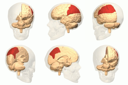
The parietal lobe is one of the four major lobes of the cerebral cortex in the brain of mammals. The parietal lobe is positioned above the temporal lobe and behind the frontal lobe and central sulcus. The parietal lobe integrates sensory information among various modalities, including spatial sense and navigation (proprioception), the main sensory receptive area for the sense of touch in the somatosensory cortex which is just posterior to the central sulcus in the postcentral gyrus, and the dorsal stream of the visual system. The major sensory inputs from the skin (touch, temperature, and pain receptors), relay through the thalamus to the parietal lobe. Several areas of the parietal lobe are important in language processing. The somatosensory cortex can be illustrated as a distorted figure – the cortical homunculus (Latin: "little man") in which the body parts are rendered according to how much of the somatosensory cortex is devoted to them. The superior parietal lobule and in ... [...More Info...] [...Related Items...] OR: [Wikipedia] [Google] [Baidu] |
Lesion
A lesion is any damage or abnormal change in the tissue of an organism, usually caused by disease or trauma. ''Lesion'' is derived from the Latin "injury". Lesions may occur in plants as well as animals. Types There is no designated classification or naming convention for lesions. Since lesions can occur anywhere in the body and the definition of a lesion is so broad, the varieties of lesions are virtually endless. Generally, lesions may be classified by their patterns, their sizes, their locations, or their causes. They can also be named after the person who discovered them. For example, Ghon lesions, which are found in the lungs of those with tuberculosis, are named after the lesion's discoverer, Anton Ghon. The characteristic skin lesions of a varicella zoster virus infection are called '' chickenpox''. Lesions of the teeth are usually called dental caries. Location Lesions are often classified by their tissue types or locations. For example, a "skin lesion" or a " bra ... [...More Info...] [...Related Items...] OR: [Wikipedia] [Google] [Baidu] |
Superficial Siderosis
Superficial hemosiderosis of the central nervous system is a disease of the brain resulting from chronic iron deposition in neuronal tissues associated with cerebrospinal fluid. This occurs via the deposition of hemosiderin in neuronal tissue, and is associated with neuronal loss, gliosis, and demyelination of neuronal cells. This disease was first discovered in 1908 by R.C. Hamill after performing an autopsy. Detection of this disease was largely post-mortem until the advent of MRI technology, which made diagnosis far easier. Superficial siderosis is largely considered a rare disease, with less than 270 total reported cases in scientific literature as of 2006, and affects people of a wide range of ages with men being approximately three times more frequently affected than women. The number of reported cases of superficial siderosis has increased with advances in MRI technology, but it remains a rare disease. Symptoms Superficial siderosis is characterized by many symptoms resultin ... [...More Info...] [...Related Items...] OR: [Wikipedia] [Google] [Baidu] |
Cerebral Amyloid Angiopathy
Cerebral amyloid angiopathy (CAA) is a form of angiopathy in which amyloid beta peptide deposits in the walls of small to medium blood vessels of the central nervous system and meninges. The term ''congophilic'' is sometimes used because the presence of the abnormal aggregations of amyloid can be demonstrated by microscopic examination of brain tissue after staining with Congo red. The amyloid material is only found in the brain and as such the disease is not related to other forms of amyloidosis. Signs and symptoms CAA is associated with brain hemorrhages, particularly microhemorrhages. Since CAA can be caused by the same amyloid protein that is associated with Alzheimer's dementia, brain bleeds are more common in people who have a diagnosis of Alzheimer's disease. However, they can also occur in those who have no history of dementia. The bleeding within the brain is usually confined to a particular lobe and this is slightly different compared to brain bleeds which occur as a ... [...More Info...] [...Related Items...] OR: [Wikipedia] [Google] [Baidu] |
White Matter
White matter refers to areas of the central nervous system (CNS) that are mainly made up of myelinated axons, also called tracts. Long thought to be passive tissue, white matter affects learning and brain functions, modulating the distribution of action potentials, acting as a relay and coordinating communication between different brain regions. White matter is named for its relatively light appearance resulting from the lipid content of myelin. However, the tissue of the freshly cut brain appears pinkish-white to the naked eye because myelin is composed largely of lipid tissue veined with capillaries. Its white color in prepared specimens is due to its usual preservation in formaldehyde. Structure White matter White matter is composed of bundles, which connect various grey matter areas (the locations of nerve cell bodies) of the brain to each other, and carry nerve impulses between neurons. Myelin acts as an insulator, which allows electrical signals to jump, rather than c ... [...More Info...] [...Related Items...] OR: [Wikipedia] [Google] [Baidu] |
Cerebral Cortex
The cerebral cortex, also known as the cerebral mantle, is the outer layer of neural tissue of the cerebrum of the brain in humans and other mammals. The cerebral cortex mostly consists of the six-layered neocortex, with just 10% consisting of allocortex. It is separated into two cortices, by the longitudinal fissure that divides the cerebrum into the left and right cerebral hemispheres. The two hemispheres are joined beneath the cortex by the corpus callosum. The cerebral cortex is the largest site of neural integration in the central nervous system. It plays a key role in attention, perception, awareness, thought, memory, language, and consciousness. The cerebral cortex is part of the brain responsible for cognition. In most mammals, apart from small mammals that have small brains, the cerebral cortex is folded, providing a greater surface area in the confined volume of the cranium. Apart from minimising brain and cranial volume, cortical folding is crucial for the brain ... [...More Info...] [...Related Items...] OR: [Wikipedia] [Google] [Baidu] |
Sulcus (neuroanatomy)
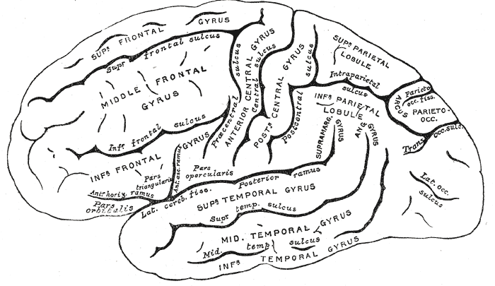
In neuroanatomy, a sulcus (Latin: "furrow", pl. ''sulci'') is a depression or groove in the cerebral cortex. It surrounds a gyrus (pl. gyri), creating the characteristic folded appearance of the brain in humans and other mammals. The larger sulci are usually called fissures. Structure Sulci, the grooves, and gyri, the folds or ridges, make up the folded surface of the cerebral cortex. Larger or deeper sulci are termed fissures, and in many cases the two terms are interchangeable. The folded cortex creates a larger surface area for the brain in humans and other mammals. When looking at the human brain, two-thirds of the surface are hidden in the grooves. The sulci and fissures are both grooves in the cortex, but they are differentiated by size. A sulcus is a shallower groove that surrounds a gyrus. A fissure is a large furrow that divides the brain into lobes and also into the two hemispheres as the longitudinal fissure. Importance of expanded surface area As the surfac ... [...More Info...] [...Related Items...] OR: [Wikipedia] [Google] [Baidu] |
Fluid-attenuated Inversion Recovery
Fluid-attenuated inversion recovery (FLAIR) is an MRI sequence with an inversion recovery set to null fluids. For example, it can be used in brain imaging to suppress cerebrospinal fluid (CSF) effects on the image, so as to bring out the periventricular hyperintense lesions, such as multiple sclerosis (MS) plaques. It was invented by Dr. Graeme Bydder. FLAIR can be used with both three-dimensional imaging (3D FLAIR) or two dimensional imaging (2D FLAIR). Technique By carefully choosing the inversion time (TI), the signal from any particular tissue can be nulled. The appropriate TI depends on the tissue via the formula: :\textrm = \ln(2) \cdot T_1,\, in other words, one should typically use a TI of around 70% of the ''T1'' value. In the case of CSF suppression, one aims for ''T1''-weighted images, which prioritize the signal of fat over that of water. Therefore, if the long TI (inversion time) is adjusted to a zero crossing point for water (none of its signal is visible), the ... [...More Info...] [...Related Items...] OR: [Wikipedia] [Google] [Baidu] |


