|
Aδ Fibers
Group A nerve fibers are one of the three classes of nerve fiber as ''generally classified'' by Erlanger and Gasser. The other two classes are the group B nerve fibers, and the group C nerve fibers. Group A are heavily myelinated, group B are moderately myelinated, and group C are unmyelinated. The other classification is a sensory grouping that uses the terms '' type Ia and type Ib'', '' type II'', ''type III'', and ''type IV'', sensory fibers. Types There are four subdivisions of group A nerve fibers: alpha (α) Aα; beta (β) Aβ; , gamma (γ) Aγ, and delta (δ) Aδ. These subdivisions have different amounts of myelination and axon thickness and therefore transmit signals at different speeds. Larger diameter axons and more myelin insulation lead to faster signal propagation. Group A nerves are found in both motor and sensory pathways. Different sensory receptors are innervated by different types of nerve fibers. Proprioceptors are innervated by type Ia, Ib and II ... [...More Info...] [...Related Items...] OR: [Wikipedia] [Google] [Baidu] |
Axon
An axon (from Greek ἄξων ''áxōn'', axis), or nerve fiber (or nerve fibre: see spelling differences), is a long, slender projection of a nerve cell, or neuron, in vertebrates, that typically conducts electrical impulses known as action potentials away from the nerve cell body. The function of the axon is to transmit information to different neurons, muscles, and glands. In certain sensory neurons (pseudounipolar neurons), such as those for touch and warmth, the axons are called afferent nerve fibers and the electrical impulse travels along these from the periphery to the cell body and from the cell body to the spinal cord along another branch of the same axon. Axon dysfunction can be the cause of many inherited and acquired neurological disorders that affect both the peripheral and central neurons. Nerve fibers are classed into three typesgroup A nerve fibers, group B nerve fibers, and group C nerve fibers. Groups A and B are myelinated, and group C are unmyelinated. ... [...More Info...] [...Related Items...] OR: [Wikipedia] [Google] [Baidu] |
Proprioceptor
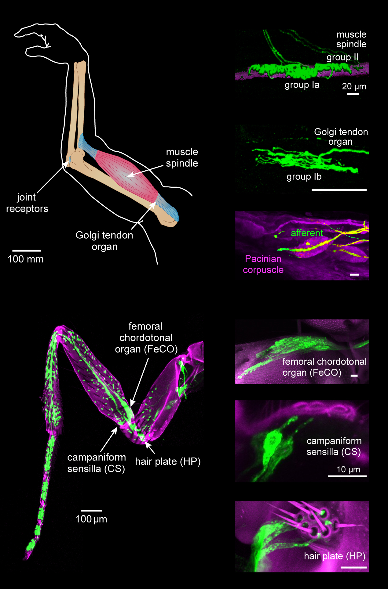
Proprioception ( ), also referred to as kinaesthesia (or kinesthesia), is the sense of self-movement, force, and body position. It is sometimes described as the "sixth sense". Proprioception is mediated by proprioceptors, mechanosensory neurons located within muscles, tendons, and joints. Most animals possess multiple subtypes of proprioceptors, which detect distinct kinematic parameters, such as joint position, movement, and load. Although all mobile animals possess proprioceptors, the structure of the sensory organs can vary across species. Proprioceptive signals are transmitted to the central nervous system, where they are integrated with information from other sensory systems, such as the visual system and the vestibular system, to create an overall representation of body position, movement, and acceleration. In many animals, sensory feedback from proprioceptors is essential for stabilizing body posture and coordinating body movement. System overview In vertebrates, limb v ... [...More Info...] [...Related Items...] OR: [Wikipedia] [Google] [Baidu] |
Neospinothalamic Tract
Nociception (also nocioception, from Latin ''nocere'' 'to harm or hurt') is the sensory nervous system's process of encoding noxious stimuli. It deals with a series of events and processes required for an organism to receive a painful stimulus, convert it to a molecular signal, and recognize and characterize the signal in order to trigger an appropriate defense response. In nociception, intense chemical (e.g., capsaicin present in Chili pepper or Cayenne pepper), mechanical (e.g., cutting, crushing), or thermal (heat and cold) stimulation of sensory neurons called nociceptors produces a signal that travels along a chain of nerve fibers via the spinal cord to the brain. Nociception triggers a variety of physiological and behavioral responses to protect the organism against an aggression and usually results in a subjective experience, or perception, of pain in sentient beings. Detection of noxious stimuli Potentially damaging mechanical, thermal, and chemical stimuli are detected ... [...More Info...] [...Related Items...] OR: [Wikipedia] [Google] [Baidu] |
Free Nerve Ending
A free nerve ending (FNE) or bare nerve ending, is an unspecialized, afferent nerve fiber sending its signal to a sensory neuron. ''Afferent'' in this case means bringing information from the body's periphery toward the brain. They function as cutaneous nociceptors and are essentially used by vertebrates to detect noxious stimuli that often result in pain. Structure Free nerve endings are unencapsulated and have no complex sensory structures. They are the most common type of nerve ending, and are most frequently found in the skin. They penetrate the dermis and end in the stratum granulosum. FNEs infiltrate the middle layers of the dermis and surround hair follicles. Types Free nerve endings have different rates of adaptation, stimulus modalities, and fiber types. Rate of adaptation Different types of FNE can be rapidly adapting, intermediate adapting, or slowly adapting. A delta type II fibers are fast-adapting while A delta type I and C fibers are slowly adapting.Rolf-Detlef ... [...More Info...] [...Related Items...] OR: [Wikipedia] [Google] [Baidu] |
Nociceptors
A nociceptor ("pain receptor" from Latin ''nocere'' 'to harm or hurt') is a sensory neuron that responds to damaging or potentially damaging stimuli by sending "possible threat" signals to the spinal cord and the brain. The brain creates the sensation of pain to direct attention to the body part, so the threat can be mitigated; this process is called nociception. History Nociceptors were discovered by Charles Scott Sherrington in 1906. In earlier centuries, scientists believed that animals were like mechanical devices that transformed the energy of sensory stimuli into motor responses. Sherrington used many different experiments to demonstrate that different types of stimulation to an afferent nerve fiber's receptive field led to different responses. Some intense stimuli trigger reflex withdrawal, certain autonomic responses, and pain. The specific receptors for these intense stimuli were called nociceptors. Location In mammals, nociceptors are found in any area of the body t ... [...More Info...] [...Related Items...] OR: [Wikipedia] [Google] [Baidu] |
Cutaneous Mechanoreceptor
A mechanoreceptor, also called mechanoceptor, is a sensory receptor that responds to mechanical pressure or distortion. Mechanoreceptors are innervated by sensory neurons that convert mechanical pressure into electrical signals that, in animals, are sent to the central nervous system. Vertebrate mechanoreceptors Cutaneous mechanoreceptors Cutaneous mechanoreceptors respond to mechanical stimuli that result from physical interaction, including pressure and vibration. They are located in the skin, like other cutaneous receptors. They are all innervated by Aβ fibers, except the mechanorecepting free nerve endings, which are innervated by Aδ fibers. Cutaneous mechanoreceptors can be categorized by what kind of sensation they perceive, by the rate of adaptation, and by morphology. Furthermore, each has a different receptive field. By sensation *The Slowly Adapting type 1 (SA1) mechanoreceptor, with the Merkel corpuscle end-organ (also known as Merkel discs) detect sustained p ... [...More Info...] [...Related Items...] OR: [Wikipedia] [Google] [Baidu] |
Muscle Spindle
Muscle spindles are stretch receptors within the body of a skeletal muscle that primarily detect changes in the length of the muscle. They convey length information to the central nervous system via afferent nerve fibers. This information can be processed by the brain as proprioception. The responses of muscle spindles to changes in length also play an important role in regulating the contraction of muscles, for example, by activating motor neurons via the stretch reflex to resist muscle stretch. The muscle spindle has both sensory and motor components. * Sensory information conveyed by primary type Ia sensory fibers which spiral around muscle fibres within the spindle, and secondary type II sensory fibers * Activation of muscle fibres within the spindle by up to a dozen gamma motor neurons and to a lesser extent by one or two beta motor neurons Structure Muscle spindles are found within the belly of a skeletal muscle. Muscle spindles are fusiform (spindle-shaped), and the spec ... [...More Info...] [...Related Items...] OR: [Wikipedia] [Google] [Baidu] |
Type II Sensory Fiber
Type II sensory fiber (group Aβ) is a type of sensory fiber, the second of the two main groups of touch receptors. The responses of different type Aβ fibers to these stimuli can be subdivided based on their adaptation properties, traditionally into rapidly adapting (RA) or slowly adapting (SA) neurons. Type II sensory fibers are slowly-adapting (SA), meaning that even when there is no change in touch, they keep respond to stimuli and fire action potentials. In the body, Type II sensory fibers belong to pseudounipolar neurons. The most notable example are neurons with Merkel cell-neurite complexes on their dendrites (sense static touch) and Ruffini endings (sense stretch on the skin and over-extension inside joints). Under pathological conditions they may become hyper-excitable leading to stimuli that would usually elicit sensations of tactile touch causing pain. These changes are in part induced by PGE2 which is produced by COX1, and type II fibers with free nerve endings ... [...More Info...] [...Related Items...] OR: [Wikipedia] [Google] [Baidu] |
Golgi Tendon Organ
The Golgi tendon organ (GTO) (also called Golgi organ, tendon organ, neurotendinous organ or neurotendinous spindle) is a proprioceptor – a type of sensory receptor that senses changes in muscle tension. It lies at the interface between a muscle and its tendon known as the musculotendinous junction also known as the myotendinous junction. It provides the sensory component of the Golgi tendon reflex. The Golgi tendon organ is one of several eponymous terms named after the Italian physician Camillo Golgi. Structure The body of the Golgi tendon organ is made up of braided strands of collagen (intrafusal fasciculi) that are less compact than elsewhere in the tendon and are encapsulated. The capsule is connected in series (along a single path) with a group of muscle fibers () at one end, and merge into the tendon proper at the other. Each capsule is about long, has a diameter of about , and is perforated by one or more afferent type Ib sensory nerve fibers ( Aɑ fiber), which ... [...More Info...] [...Related Items...] OR: [Wikipedia] [Google] [Baidu] |
Proprioception
Proprioception ( ), also referred to as kinaesthesia (or kinesthesia), is the sense of self-movement, force, and body position. It is sometimes described as the "sixth sense". Proprioception is mediated by proprioceptors, mechanosensory neurons located within muscles, tendons, and joints. Most animals possess multiple subtypes of proprioceptors, which detect distinct kinematic parameters, such as joint position, movement, and load. Although all mobile animals possess proprioceptors, the structure of the sensory organs can vary across species. Proprioceptive signals are transmitted to the central nervous system, where they are integrated with information from other sensory systems, such as the visual system and the vestibular system, to create an overall representation of body position, movement, and acceleration. In many animals, sensory feedback from proprioceptors is essential for stabilizing body posture and coordinating body movement. System overview In vertebrates, limb ve ... [...More Info...] [...Related Items...] OR: [Wikipedia] [Google] [Baidu] |
Type Ia Sensory Fiber
A type Ia sensory fiber, or a primary afferent fiber is a type of afferent nerve fiber. It is the sensory fiber of a stretch receptor called the muscle spindle found in muscles, which constantly monitors the rate at which a muscle stretch changes. The information carried by type Ia fibers contributes to the sense of proprioception. Function of muscle spindles For the body to keep moving properly and with finesse, the nervous system has to have a constant input of sensory data coming from areas such as the muscles and joints. In order to receive a continuous stream of sensory data, the body has developed special sensory receptors called proprioceptors. Muscle spindles are a type of proprioceptor, and they are found inside the muscle itself. They lie parallel with the contractile fibers. This gives them the ability to monitor muscle length with precision. Types of sensory fibers This change in length of the spindle is transduced (transformed into electric membrane potentials ... [...More Info...] [...Related Items...] OR: [Wikipedia] [Google] [Baidu] |
Sensory Receptor
Sensory neurons, also known as afferent neurons, are neurons in the nervous system, that convert a specific type of stimulus, via their receptors, into action potentials or graded potentials. This process is called sensory transduction. The cell bodies of the sensory neurons are located in the dorsal ganglia of the spinal cord. The sensory information travels on the afferent nerve fibers in a sensory nerve, to the brain via the spinal cord. The stimulus can come from ''exteroreceptors'' outside the body, for example those that detect light and sound, or from ''interoreceptors'' inside the body, for example those that are responsive to blood pressure or the sense of body position. Types and function Different types of sensory neurons have different sensory receptors that respond to different kinds of stimuli. There are at least six external and two internal sensory receptors: External receptors External receptors that respond to stimuli from outside the body are called ex ... [...More Info...] [...Related Items...] OR: [Wikipedia] [Google] [Baidu] |


