|
Astroblastoma
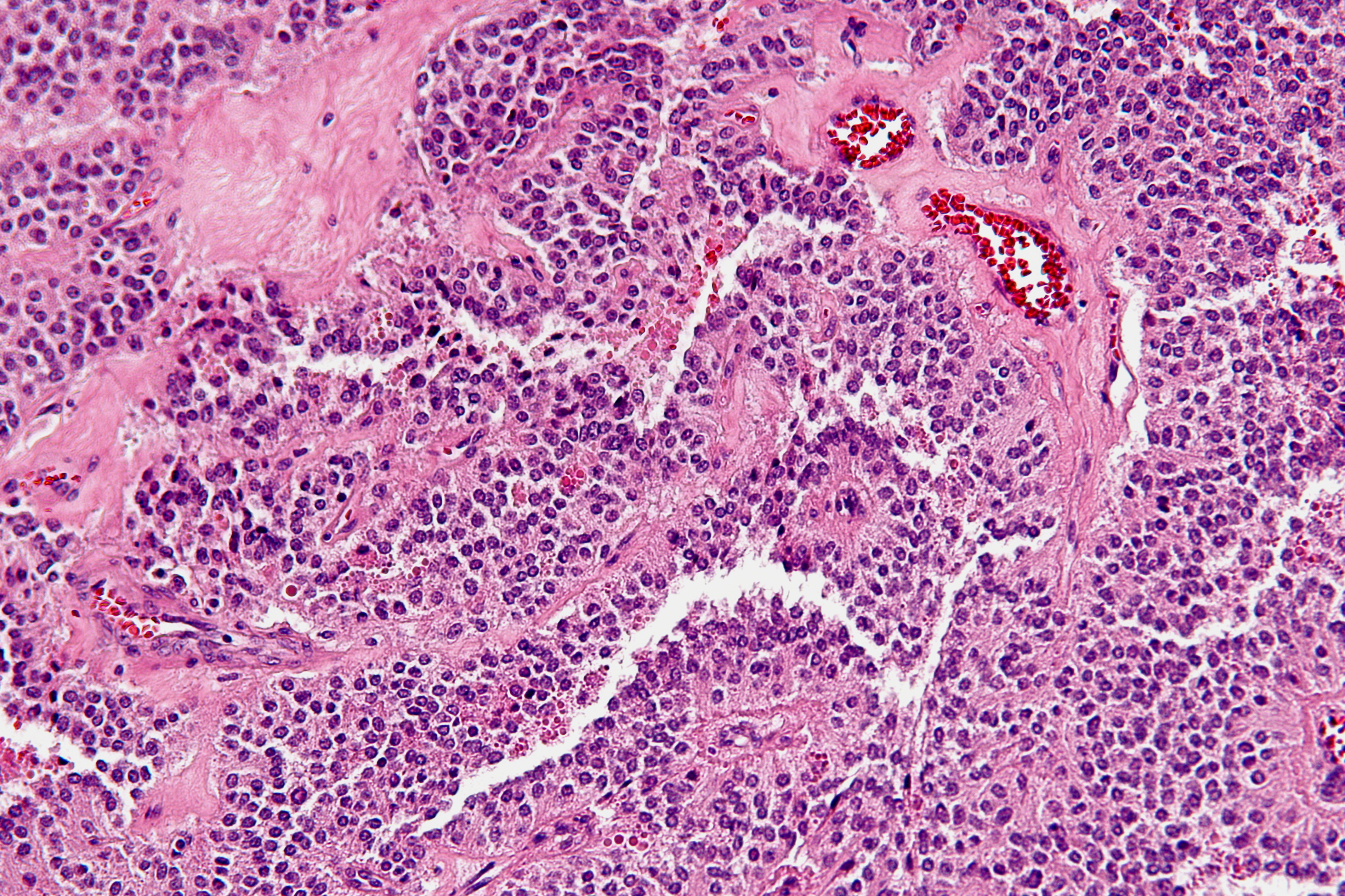
Astroblastoma is a rare glial tumor derived from the astroblast, a type of cell that closely resembles spongioblastoma and astrocytes. Astroblastoma cells are most likely found in the supratentorial region of the brain that houses the cerebrum, an area responsible for all voluntary movements in the body. It also occurs significantly in the frontal lobe, parietal lobe, and temporal lobe, areas where movement, language creation, memory perception, and environmental surroundings are expressed. These tumors can be present in major brain areas not associated with the main cerebral hemispheres, including the cerebellum, optic nerve, cauda equina, hypothalamus, and brain stem. The most defining physical symptom of astroblastoma, regardless of location, is elevated intracranial pressure, occurring when cerebrospinal fluid in the subarachnoid space exhibits heavy pressure and decreased blood flow, resulting in throbbing headache or nausea for the patient. Despite widespread localization ... [...More Info...] [...Related Items...] OR: [Wikipedia] [Google] [Baidu] |
Micrograph
A micrograph or photomicrograph is a photograph or digital image taken through a microscope or similar device to show a magnified image of an object. This is opposed to a macrograph or photomacrograph, an image which is also taken on a microscope but is only slightly magnified, usually less than 10 times. Micrography is the practice or art of using microscopes to make photographs. A micrograph contains extensive details of microstructure. A wealth of information can be obtained from a simple micrograph like behavior of the material under different conditions, the phases found in the system, failure analysis, grain size estimation, elemental analysis and so on. Micrographs are widely used in all fields of microscopy. Types Photomicrograph A light micrograph or photomicrograph is a micrograph prepared using an optical microscope, a process referred to as ''photomicroscopy''. At a basic level, photomicroscopy may be performed simply by connecting a camera to a microscope, th ... [...More Info...] [...Related Items...] OR: [Wikipedia] [Google] [Baidu] |
Epithelioid
Epithelioid may refer to: * Epithelioid cell, a cell that resembles epithelial cells * Epithelioid sarcoma, a soft tissue tumour * Epithelioid hemangioendothelioma, a vascular tumour occurring in the lining of blood vessels * Epithelioid blue nevus, a melanocytic nevus * Epithelioid sarcoma-like hemangioendothelioma, a group of vascular neoplasms * Epithelioid and spindle-cell nevus, a benign melanocytic lesion affecting the epidermis and dermis * Epithelioid hemangioma Epithelioid may refer to: * Epithelioid cell, a cell that resembles epithelial cells * Epithelioid sarcoma, a soft tissue tumour * Epithelioid hemangioendothelioma, a vascular tumour occurring in the lining of blood vessels * Epithelioid blue nevus ... * Epithelioid cell histiocytoma, a skin condition similar to dermatofibroma External links {{disambig ... [...More Info...] [...Related Items...] OR: [Wikipedia] [Google] [Baidu] |
Midline
In typography, the mean line is the imaginary line at the top of the x-height. upright 2.0, alt=A diagram showing the line terms used in typography Round glyphs will tend to break ( overshoot) the mean line slightly in many typefaces, since this is aesthetically more pleasing, otherwise curved letters such as ''a'', ''c'', ''e'', ''m'', ''n'', ''o'', ''r'', ''s'', and ''u'' will appear visually smaller than flat-topped (or bottomed) characters of equal height, due to an optical illusion Within visual perception, an optical illusion (also called a visual illusion) is an illusion caused by the visual system and characterized by a visual perception, percept that arguably appears to differ from reality. Illusions come in a wide v .... References External links * Page 31The Complete Manual of Typography: A Guide to Setting Perfect Type, Second Editionby James Felici {{DEFAULTSORT:Mean line Typography ... [...More Info...] [...Related Items...] OR: [Wikipedia] [Google] [Baidu] |
Fourth Ventricle
The fourth ventricle is one of the four connected fluid-filled cavities within the human brain. These cavities, known collectively as the ventricular system, consist of the left and right lateral ventricles, the third ventricle, and the fourth ventricle. The fourth ventricle extends from the cerebral aqueduct (''aqueduct of Sylvius'') to the obex, and is filled with cerebrospinal fluid (CSF). The fourth ventricle has a characteristic diamond shape in cross-sections of the human brain. It is located within the pons or in the upper part of the medulla oblongata. CSF entering the fourth ventricle through the cerebral aqueduct can exit to the subarachnoid space of the spinal cord through two lateral apertures and a single, midline median aperture. Boundaries The fourth ventricle has a roof at its ''upper'' (posterior) surface and a floor at its ''lower'' (anterior) surface, and side walls formed by the cerebellar peduncles (nerve bundles joining the structure on the posterior sid ... [...More Info...] [...Related Items...] OR: [Wikipedia] [Google] [Baidu] |
Posterior Inferior Cerebellar Artery
The posterior inferior cerebellar artery (PICA) is the largest branch of the vertebral artery. It is one of the three main arteries that supply blood to the cerebellum, a part of the brain. Blockage of the posterior inferior cerebellar artery can result in a type of stroke called lateral medullary syndrome. Course It winds backward around the upper part of the medulla oblongata, passing between the origins of the vagus nerve and the accessory nerve, over the inferior cerebellar peduncle to the undersurface of the cerebellum, where it divides into two branches. The medial branch continues backward to the notch between the two hemispheres of the cerebellum; while the lateral supplies the under surface of the cerebellum, as far as its lateral border, where it anastomoses with the anterior inferior cerebellar and the superior cerebellar branches of the basilar artery. Branches from this artery supply the choroid plexus of the fourth ventricle. Clinical significance A disrupted blo ... [...More Info...] [...Related Items...] OR: [Wikipedia] [Google] [Baidu] |
Computerized Tomography
A computed tomography scan (CT scan; formerly called computed axial tomography scan or CAT scan) is a medical imaging technique used to obtain detailed internal images of the body. The personnel that perform CT scans are called radiographers or radiology technologists. CT scanners use a rotating X-ray tube and a row of detectors placed in a gantry to measure X-ray attenuations by different tissues inside the body. The multiple X-ray measurements taken from different angles are then processed on a computer using tomographic reconstruction algorithms to produce tomographic (cross-sectional) images (virtual "slices") of a body. CT scans can be used in patients with metallic implants or pacemakers, for whom magnetic resonance imaging (MRI) is contraindicated. Since its development in the 1970s, CT scanning has proven to be a versatile imaging technique. While CT is most prominently used in medical diagnosis, it can also be used to form images of non-living objects. The 1979 Nob ... [...More Info...] [...Related Items...] OR: [Wikipedia] [Google] [Baidu] |
Calcification
Calcification is the accumulation of calcium salts in a body tissue. It normally occurs in the formation of bone, but calcium can be deposited abnormally in soft tissue,Miller, J. D. Cardiovascular calcification: Orbicular origins. ''Nature Materials'' 12, 476-478 (2013). causing it to harden. Calcifications may be classified on whether there is mineral balance or not, and the location of the calcification. Calcification may also refer to the processes of normal mineral deposition in biological systems, such as the formation of stromatolites or mollusc shells (see Biomineralization). Signs and symptoms Calcification can manifest itself in many ways in the body depending on the location. In the pulpal structure of a tooth, calcification often presents asymptomatically, and is diagnosed as an incidental finding during radiographic interpretation. Individual teeth with calcified pulp will typically respond negatively to vitality testing; teeth with calcified pulp often lack sen ... [...More Info...] [...Related Items...] OR: [Wikipedia] [Google] [Baidu] |
Hyalinization
A hyaline substance is one with a glassy appearance. The word is derived from el, ὑάλινος, translit=hyálinos, lit=transparent, and el, ὕαλος, translit=hýalos, lit=crystal, glass, label=none. Histopathology Hyaline cartilage is named after its glassy appearance on fresh gross pathology. On light microscopy of H&E stained slides, the extracellular matrix of hyaline cartilage looks homogeneously pink, and the term "hyaline" is used to describe similarly homogeneously pink material besides the cartilage. Hyaline material is usually acellular and proteinaceous. For example, arterial hyaline is seen in aging, high blood pressure, diabetes mellitus and in association with some drugs (e.g. calcineurin inhibitors). It is bright pink with PAS staining. Ichthyology and entomology In ichthyology and entomology, ''hyaline'' denotes a colorless, transparent substance, such as unpigmented fins of fishes or clear insect wings. Resh, Vincent H. and R. T. Cardé, Eds. Encyclo ... [...More Info...] [...Related Items...] OR: [Wikipedia] [Google] [Baidu] |
Fibrosis
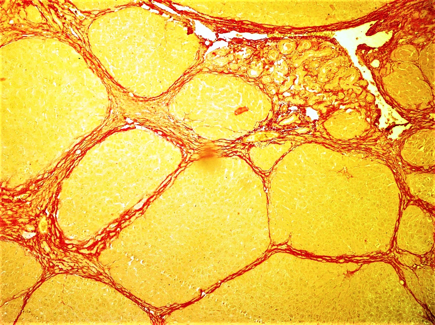
Fibrosis, also known as fibrotic scarring, is a pathological wound healing in which connective tissue replaces normal parenchymal tissue to the extent that it goes unchecked, leading to considerable tissue remodelling and the formation of permanent scar tissue. Repeated injuries, chronic inflammation and repair are susceptible to fibrosis where an accidental excessive accumulation of extracellular matrix components, such as the collagen is produced by fibroblasts, leading to the formation of a permanent fibrotic scar. In response to injury, this is called scarring, and if fibrosis arises from a single cell line, this is called a fibroma. Physiologically, fibrosis acts to deposit connective tissue, which can interfere with or totally inhibit the normal architecture and function of the underlying organ or tissue. Fibrosis can be used to describe the pathological state of excess deposition of fibrous tissue, as well as the process of connective tissue deposition in healing. Define ... [...More Info...] [...Related Items...] OR: [Wikipedia] [Google] [Baidu] |
Cerebral Hemispheres
The vertebrate cerebrum (brain) is formed by two cerebral hemispheres that are separated by a groove, the longitudinal fissure. The brain can thus be described as being divided into left and right cerebral hemispheres. Each of these hemispheres has an outer layer of grey matter, the cerebral cortex, that is supported by an inner layer of white matter. In eutherian (placental) mammals, the hemispheres are linked by the corpus callosum, a very large bundle of nerve fibers. Smaller commissures, including the anterior commissure, the posterior commissure and the fornix, also join the hemispheres and these are also present in other vertebrates. These commissures transfer information between the two hemispheres to coordinate localized functions. There are three known poles of the cerebral hemispheres: the ''occipital pole'', the ''frontal pole'', and the ''temporal pole''. The central sulcus is a prominent fissure which separates the parietal lobe from the frontal lobe and the prim ... [...More Info...] [...Related Items...] OR: [Wikipedia] [Google] [Baidu] |
Solid Pseudopapillary Tumor
A solid pseudopapillary tumour is a low-grade malignant neoplasm of the pancreas of :wikt:papilla, papillary architecture that typically afflicts young women. Signs and symptoms Solid pseudopapillary tumours are often asymptomatic and are identified incidentally on imaging performed for unrelated reasons. Less often, they may cause abdominal pain. Solid pseudopapillary tumours tend to occur in women, and most often present in the third decade of life. Anatomy Gross morphology Solid pseudopapillary tumours are typically round, well-demarcated, measuring 2–17 cm in diameter (average 8 cm), with solid and cystic areas with hemorrhage on cut sections. Histomorphology Solid pseudopapillary tumours consist of solid sheets of cells that are focally dyscohesive. The cells in the lesion usually have uniform nucleus (cell), nuclei with occasional nuclear grooves, eosinophilic or clear cytoplasm and Periodic acid-Schiff stain, PAS positive eosinophilic intracytoplasmic globule ... [...More Info...] [...Related Items...] OR: [Wikipedia] [Google] [Baidu] |
Rosette (design)
A rosette is a round, stylized flower design. Origin The rosette derives from the natural shape of the botanical rosette, formed by leaves radiating out from the stem of a plant and visible even after the flowers have withered. History The rosette design is used extensively in sculptural objects from antiquity, appearing in Mesopotamia, and in funeral steles' decoration in Ancient Greece. The rosette was another important symbol of Ishtar which had originally belonged to Inanna along with the Star of Ishtar. It was adopted later in Romaneseque and Renaissance architecture, and also common in the art of Central Asia, spreading as far as India where it is used as a decorative motif in Greco-Buddhist art. Ancient origins One of the earliest appearances of the rosette in ancient art is in early fourth millennium BC Egypt. Another early Mediterranean occurrence of the rosette design derives from Minoan Crete; Among other places, the design appears on the Phaistos Disc, recove ... [...More Info...] [...Related Items...] OR: [Wikipedia] [Google] [Baidu] |


_-_inferiror_view.png)
