|
Arcuate Ligament (other)
The arcuate ligament may refer to: * Inferior pubic ligament (arcuate ligament of the pubis). * Arcuate popliteal ligament. * Arcuate ligaments of the diaphragm: ** Median arcuate ligament ** Medial arcuate ligament ** Lateral arcuate ligament The lateral arcuate ligament (also lateral lumbocostal arch and external arcuate ligament) is a ligament under the diaphragm that arches across the upper part of the quadratus lumborum muscle. It is traversed by the subcostal nerve, artery and vein ... {{DEFAULTSORT:Arcuate Ligament Ligaments ... [...More Info...] [...Related Items...] OR: [Wikipedia] [Google] [Baidu] |
Inferior Pubic Ligament
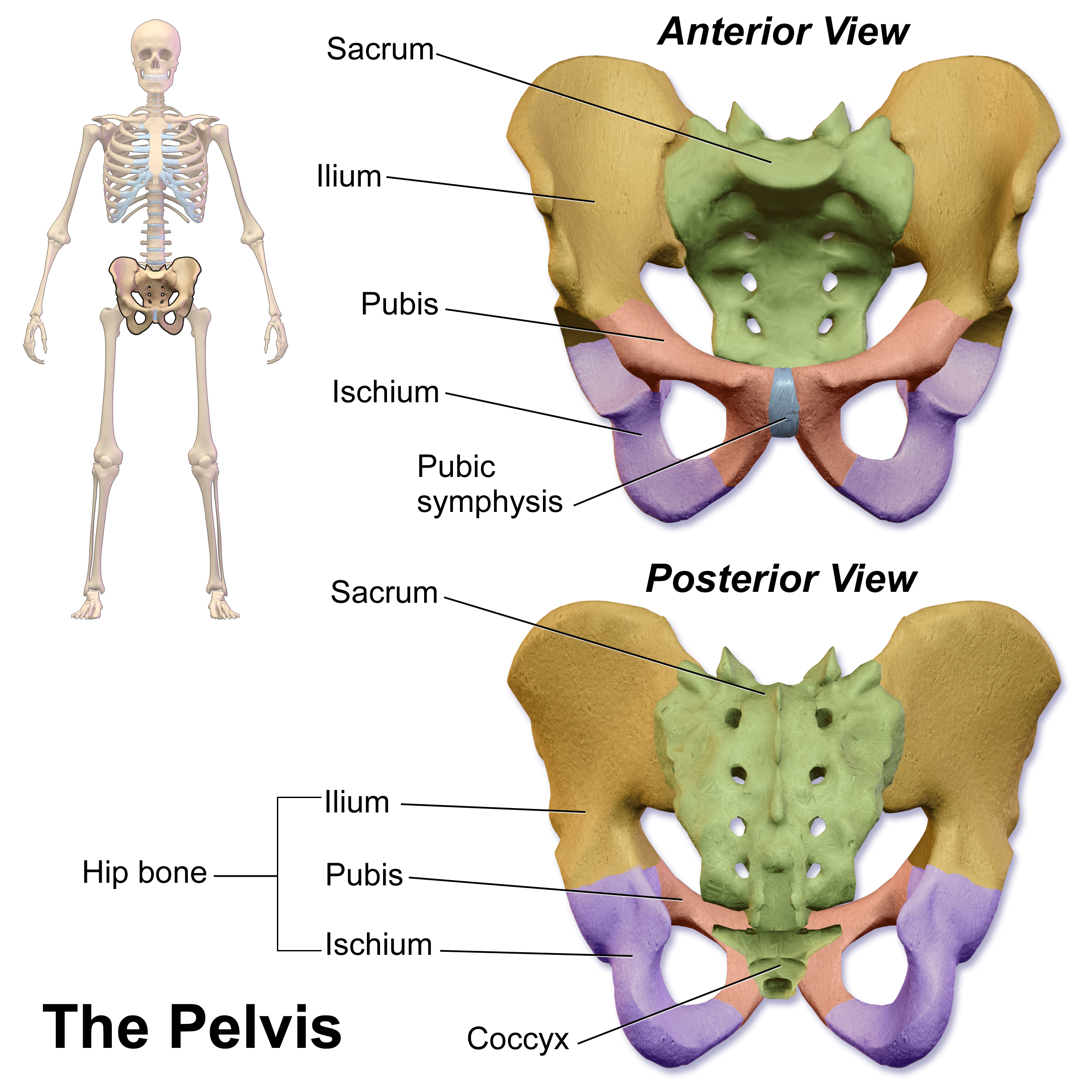
The pubic symphysis is a secondary cartilaginous joint between the left and right superior rami of the pubis of the hip bones. It is in front of and below the urinary bladder. In males, the suspensory ligament of the penis attaches to the pubic symphysis. In females, the pubic symphysis is close to the clitoris. In most adults it can be moved roughly 2 mm and with 1 degree rotation. This increases for women at the time of childbirth. The name comes from the Greek word ''symphysis'', meaning 'growing together'. Structure The pubic symphysis is a nonsynovial amphiarthrodial joint. The width of the pubic symphysis at the front is 3–5 mm greater than its width at the back. This joint is connected by fibrocartilage and may contain a fluid-filled cavity; the center is avascular, possibly due to the nature of the compressive forces passing through this joint, which may lead to harmful vascular disease. The ends of both pubic bones are covered by a thin layer of hyaline ... [...More Info...] [...Related Items...] OR: [Wikipedia] [Google] [Baidu] |
Arcuate Popliteal Ligament
The arcuate popliteal ligament is an Y-shaped extracapsular ligament of the knee. It is formed as a thickening of the posterior fibres of the joint capsule of the knee. It has its origin at the posterior aspect of the head of the fibula. It has two insertions: the medial limb arches superficially over the tendon of the popliteus muscle to blend with the oblique popliteal ligament; the lateral limb passes to the lateral epicondyle of the femur (accompanied by the popliteus muscle tendon) to blend there with the lateral head of the gastrocnemius muscle The gastrocnemius muscle (plural ''gastrocnemii'') is a superficial two-headed muscle that is in the back part of the lower leg of humans. It runs from its two heads just above the knee to the heel, a three joint muscle (knee, ankle and subtala .... References External links * () * - "Major Joints of the Lower Extremity: Knee Joint" Ligaments of the lower limb {{ligament-stub ... [...More Info...] [...Related Items...] OR: [Wikipedia] [Google] [Baidu] |
Median Arcuate Ligament
The median arcuate ligament is a ligament under the diaphragm that connects the right and left crura of diaphragm. Structure The median arcuate ligament is formed by the right and left crura of the diaphragm. The crura connect to form an arch, behind which is the aortic hiatus, through which pass the aorta, the azygos vein, and the thoracic duct. Variation In between 10% and 24% of people, the median arcuate ligament occurs very low. Clinical significance Compression of celiac artery and celiac ganglia by the median arcuate ligament can lead to the median arcuate ligament syndrome, which is characterized by abdominal pain, weight loss, and an epigastric bruit. See also * Medial arcuate ligament * Lateral arcuate ligament The lateral arcuate ligament (also lateral lumbocostal arch and external arcuate ligament) is a ligament under the diaphragm that arches across the upper part of the quadratus lumborum muscle. It is traversed by the subcostal nerve, artery and vein .. ... [...More Info...] [...Related Items...] OR: [Wikipedia] [Google] [Baidu] |
Medial Arcuate Ligament
The medial arcuate ligament (also medial lumbocostal arch and internal arcuate ligament) is a tendinous fascia that arches over the psoas major muscle as it passes posterior the diaphragm. Structure The medial arcuate ligament is an arch in the fascia covering the upper part of the psoas major. It is attached to the side of the body of the first or second lumbar vertebra, laterally, it is fixed to the front of the transverse process of the first and, sometimes also, to that of the second lumbar vertebra. It lies between the lateral arcuate ligament and the midline median arcuate ligament. The sympathetic chain enters the abdomen by passing deep into this ligament of the diaphragm. This is in contrast to the parasympathetic Vagus nerve which passes through the esophageal hiatus. See also * Lateral arcuate ligament * Median arcuate ligament The median arcuate ligament is a ligament under the diaphragm that connects the right and left crura of diaphragm. Structure The medi ... [...More Info...] [...Related Items...] OR: [Wikipedia] [Google] [Baidu] |
Lateral Arcuate Ligament
The lateral arcuate ligament (also lateral lumbocostal arch and external arcuate ligament) is a ligament under the diaphragm that arches across the upper part of the quadratus lumborum muscle. It is traversed by the subcostal nerve, artery and vein. Structure The lateral arcuate ligament runs from the front of the transverse process of the first lumbar vertebra, and, laterally, to the tip and lower margin of the twelfth rib. It forms an arch over the quadratus lumborum muscle. Variations The lateral arcuate ligament is commonly described in anatomy textbooks as attaching at the first lumbar vertebra (L1). However, other instances have been found in cadaver studies with attachments at either the second (L2) or third (L3) lumbar vertebra. In around 5% of people, inferolateral extensions of the lateral arcuate ligaments, such as thickened nodular areas, are found adjacent to the lateral diaphragmatic surface which can be visualized with computed tomography (CT) scans. History The l ... [...More Info...] [...Related Items...] OR: [Wikipedia] [Google] [Baidu] |
