|
Arches Of The Foot
The arches of the foot, formed by the tarsal and metatarsal bones, strengthened by ligaments and tendons, allow the foot to support the weight of the body in the erect posture with the least weight. They are categorized as longitudinal and transverse arches. Structure Longitudinal arches The longitudinal arches of the foot can be divided into medial and lateral arches. Medial arch The medial arch is higher than the lateral longitudinal arch. It is made up by the calcaneus, the talus, the navicular, the three cuneiforms (medial, intermediate, and lateral), and the first, second, and third metatarsals. Its summit is at the superior articular surface of the talus, and its two extremities or piers, on which it rests in standing, are the tuberosity on the plantar surface of the calcaneus posteriorly and the heads of the first, second, and third metatarsal bones anteriorly. The chief characteristic of this arch is its elasticity, due to its height and to the number of ... [...More Info...] [...Related Items...] OR: [Wikipedia] [Google] [Baidu] |
Tarsus (skeleton)
In the human body, the tarsus (: tarsi) is a cluster of seven articulating bones in each foot situated between the lower end of the tibia and the fibula of the lower leg and the metatarsus. It is made up of the midfoot (Cuboid bone, cuboid, medial, intermediate, and lateral cuneiform bone, cuneiform, and navicular) and hindfoot (Talus bone, talus and calcaneus). The tarsus articulates with the bones of the metatarsus, which in turn articulate with the proximal phalanges of the toes. The joint between the tibia and fibula above and the tarsus below is referred to as the ankle, ankle joint proper. In humans the largest bone in the tarsus is the calcaneus, which is the weight-bearing bone within the heel of the foot. Human anatomy Bones The talus bone or ankle bone is connected superiorly to the two bones of the lower leg, the tibia and fibula, to form the ankle, ankle joint or talocrural joint; inferiorly, at the subtalar joint, to the calcaneus or heel bone. Together, the ... [...More Info...] [...Related Items...] OR: [Wikipedia] [Google] [Baidu] |
Plantar Aponeurosis
The plantar fascia or plantar aponeurosis is the thick connective tissue aponeurosis which supports the arch on the bottom (plantar side) of the foot. Recent studies suggest that the plantar fascia is actually an aponeurosis rather than true fascia. It runs from the tuberosity of the calcaneus (heel bone) forward to the heads of the metatarsal bones (the bone between each toe and the bones of the mid-foot). Anatomy The plantar fascia is the thick central portion of the fascia investing the plantar muscles. It extends between the medial process of the and the proximal phalanges of the toes. It provides some attachment to the flexor muscles of the toes. Distally, the plantar fascia becomes continuous with the fibrous sheaths enveloping the flexor tendons passing to the toes. At the anterior extremity of the sole inferior to the heads of the metatarsal bones the plantar aponeurosis forms the superficial transverse metatarsal ligament. Structure The plantar fascia is made u ... [...More Info...] [...Related Items...] OR: [Wikipedia] [Google] [Baidu] |
Estrogen
Estrogen (also spelled oestrogen in British English; see spelling differences) is a category of sex hormone responsible for the development and regulation of the female reproductive system and secondary sex characteristics. There are three major endogenous estrogens that have estrogenic hormonal activity: estrone (E1), estradiol (E2), and estriol (E3). Estradiol, an estrane, is the most potent and prevalent. Another estrogen called estetrol (E4) is produced only during pregnancy. Estrogens are synthesized in all vertebrates and some insects. Quantitatively, estrogens circulate at lower levels than androgens in both men and women. While estrogen levels are significantly lower in males than in females, estrogens nevertheless have important physiological roles in males. Like all steroid hormones, estrogens readily diffuse across the cell membrane. Once inside the cell, they bind to and activate estrogen receptors (ERs) which in turn modulate the expression of many ... [...More Info...] [...Related Items...] OR: [Wikipedia] [Google] [Baidu] |
Pregnancy
Pregnancy is the time during which one or more offspring gestation, gestates inside a woman's uterus. A multiple birth, multiple pregnancy involves more than one offspring, such as with twins. Conception (biology), Conception usually occurs following sexual intercourse, vaginal intercourse, but can also occur through assisted reproductive technology procedures. A pregnancy may end in a Live birth (human), live birth, a miscarriage, an Abortion#Induced, induced abortion, or a stillbirth. Childbirth typically occurs around 40 weeks from the start of the Menstruation#Onset and frequency, last menstrual period (LMP), a span known as the Gestational age (obstetrics), ''gestational age''; this is just over nine months. Counting by Human fertilization#Fertilization age, ''fertilization age'', the length is about 38 weeks. Implantation (embryology), Implantation occurs on average 8–9 days after Human fertilization, fertilization. An ''embryo'' is the term for the deve ... [...More Info...] [...Related Items...] OR: [Wikipedia] [Google] [Baidu] |
Soft Tissue
Soft tissue connective tissue, connects and surrounds or supports internal organs and bones, and includes muscle, tendons, ligaments, Adipose tissue, fat, fibrous tissue, Lymphatic vessel, lymph and blood vessels, fasciae, and synovial membranes. Soft tissue is Tissue (biology), tissue in the body that is not hard tissue, hardened by the processes of ossification or calcification such as bones and teeth. It is sometimes defined by what it is not – such as "nonepithelial, extraskeletal mesenchyme exclusive of the reticuloendothelial system and glia". Composition The characteristic substances inside the extracellular matrix of soft tissue are the collagen, elastin and ground substance. Normally the soft tissue is very hydrated because of the ground substance. The fibroblasts are the most common cell responsible for the production of soft tissues' fibers and ground substance. Variations of fibroblasts, like chondroblasts, may also produce these substances. Mechanical character ... [...More Info...] [...Related Items...] OR: [Wikipedia] [Google] [Baidu] |
Adductor Hallucis
The Adductor hallucis (adductor obliquus hallucis) arises by two heads—oblique and transverse and is responsible for adducting the big toe. It has two heads, both are innervated by the lateral plantar nerve. Structure Oblique head The ''oblique head'' is a large, thick, fleshy mass, crossing the foot obliquely and occupying the hollow space under the first, second, third and fourth metatarsal bones. It arises from the bases of the second, third, and fourth metatarsal bones, and from the sheath of the tendon of the Peroneus longus, and is inserted, together with the lateral portion of the flexor hallucis brevis, into the lateral side of the base of the first phalanx of the great toe. Transverse head The ''transverse head'' (''Transversus pedis'') is a narrow, flat fasciculus which arises from the plantar metatarsophalangeal ligaments of the third, fourth, and fifth toes (sometimes only from the third and fourth), and from the transverse ligament of the metatarsals. It is insert ... [...More Info...] [...Related Items...] OR: [Wikipedia] [Google] [Baidu] |
Transverse Arch
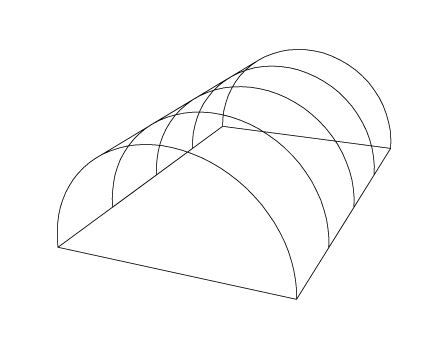
In architecture, a transverse arch is an arch in a vaulted building that goes across the barrel vault. A series of transverse arches sitting on tops of the columns on the sides of the nave was typical in the churches of Romanesque architecture (common since Carolingian times). By analogy, the term is also used to describe the transverse ribs of a groined vault and for any crosswise arch in modern buildings. An arc that goes in transverse direction, but carries an exposed wall on top, dividing the vault into compartments, is called a diaphragm arch. In the historical buildings, the transverse arches provide support for purlins and roof ridge beams. They also subdivide the nave into bays. The springings of the arch are typically pinned to supports using wooden or steel ties, but the bulk of lateral thrust is terminated in the abutments. File:Kerkplattegrond gordelboog en travee.png, The transverse arches are highlighted in green on a plan of a church File:Voûte en berceau C ... [...More Info...] [...Related Items...] OR: [Wikipedia] [Google] [Baidu] |
Foot Care And Shoe Fitting-Fig5 Cross Section Of Feet Showing Metatarsal Bones Forming Anterior Arch
The foot (: feet) is an anatomical structure found in many vertebrates. It is the terminal portion of a limb which bears weight and allows locomotion. In many animals with feet, the foot is an organ at the terminal part of the leg made up of one or more segments or bones, generally including claws and/or nails. Etymology The word "foot", in the sense of meaning the "terminal part of the leg of a vertebrate animal" comes from Old English ''fot'', from Proto-Germanic *''fot'' (source also of Old Frisian ''fot'', Old Saxon ''fot'', Old Norse ''fotr'', Danish ''fod'', Swedish ''fot'', Dutch ''voet'', Old High German ''fuoz'', German ''Fuß'', Gothic ''fotus'', all meaning "foot"), from PIE root *''ped-'' "foot". The plural form ''feet'' is an instance of i-mutation. Structure The human foot is a strong and complex mechanical structure containing 26 bones, 33 joints (20 of which are actively articulated), and more than a hundred muscles, tendons, and ligaments.Podiatry Channel, ... [...More Info...] [...Related Items...] OR: [Wikipedia] [Google] [Baidu] |
Little Toe
Toes are the digits of the foot of a tetrapod. Animal species such as cats that walk on their toes are described as being ''digitigrade''. Humans, and other animals that walk on the soles of their feet, are described as being ''plantigrade''; ''unguligrade'' animals are those that walk on hooves at the tips of their toes. Structure There are normally five toes present on each human foot. Each toe consists of three phalanx bones, the proximal, middle, and distal, with the exception of the big toe (). For a minority of people, the little toe also is missing a middle bone. The hallux only contains two phalanx bones, the proximal and distal. The joints between each phalanx are the interphalangeal joints. The proximal phalanx bone of each toe articulates with the metatarsal bone of the foot at the metatarsophalangeal joint. Each toe is surrounded by skin, and present on all five toes is a toenail. The toes are, from medial to lateral: * the first toe, also known as the hal ... [...More Info...] [...Related Items...] OR: [Wikipedia] [Google] [Baidu] |
Extensor Tendons
In anatomy, extension is a movement of a joint that increases the angle between two bones or body surfaces at a joint. Extension usually results in straightening of the bones or body surfaces involved. For example, extension is produced by extending the flexed (bent) elbow. Straightening of the arm would require extension at the elbow joint. If the head is tilted all the way back, the neck is said to be extended. Extensor muscles Upper limb *of arm at shoulder **Axilla and shoulder ***Latissimus dorsi *** Posterior fibres of deltoid ***Teres major *of forearm at elbow **Posterior compartment of the arm ***Triceps brachii *** Anconeus *of hand at wrist **Posterior compartment of the forearm *** Extensor carpi radialis longus ***Extensor carpi radialis brevis ***Extensor carpi ulnaris *** Extensor digitorum *of phalanges, at all joints **Posterior compartment of the forearm *** Extensor digitorum ***Extensor digiti minimi (little finger only) ***Extensor indicis (index finger only ... [...More Info...] [...Related Items...] OR: [Wikipedia] [Google] [Baidu] |
Plantar Calcaneocuboid
Standard anatomical terms of location are used to describe unambiguously the anatomy of humans and other animals. The terms, typically derived from Latin or Greek roots, describe something in its standard anatomical position. This position provides a definition of what is at the front ("anterior"), behind ("posterior") and so on. As part of defining and describing terms, the body is described through the use of anatomical planes and axes. The meaning of terms that are used can change depending on whether a vertebrate is a biped or a quadruped, due to the difference in the neuraxis, or if an invertebrate is a non-bilaterian. A non-bilaterian has no anterior or posterior surface for example but can still have a descriptor used such as proximal or distal in relation to a body part that is nearest to, or furthest from its middle. International organisations have determined vocabularies that are often used as standards for subdisciplines of anatomy. For example, ''Terminologia Anato ... [...More Info...] [...Related Items...] OR: [Wikipedia] [Google] [Baidu] |
Long Plantar
The long plantar ligament (long calcaneocuboid ligament; superficial long plantar ligament) is a long ligament on the underside of the foot that connects the calcaneus with the 2nd to 5th metatarsal. Structure The long plantar ligament is the longest of all the ligaments of the tarsus. It is attached behind to the plantar surface of the calcaneus in front of the tuberosity, and in front to the tuberosity on the plantar surface of the cuboid bone, the more superficial fibers being continued forward to the bases of the second, third, and fourth metatarsal bones. This ligament converts the groove on the plantar surface of the cuboid into a canal for the tendon of the fibularis longus. Deep to this ligament is the short plantar ligament. The long plantar ligament separates the two heads of the quadratus plantae muscle. See also * Plantar calcaneonavicular ligament The plantar calcaneonavicular ligament (also known as the spring ligament or spring ligament complex) is a compl ... [...More Info...] [...Related Items...] OR: [Wikipedia] [Google] [Baidu] |


