|
Anterior Clinoid Process
In the sphenoid bone, the posterior border, smooth and rounded, is received into the lateral fissure of the brain; the medial end of this border forms the anterior clinoid process, which gives attachment to the tentorium cerebelli; it is sometimes joined to the middle clinoid process by a spicule of bone, and when this occurs the termination of the groove for the internal carotid artery is converted into a foramen (carotico-clinoid). Etymology The anterior and posterior clinoid processes surround the sella turcica like the four corners of a four poster bed. Cline is Greek for bed. –oid, as usual, indicates a similarity to. The term may also come from the Greek root ''klinein'' or the Latin ''clinare'', both meaning "sloped" as in "inclined". Additional images Image:Anterior clinoid process.jpg, Anterior clinoid process Petroclinoid ligament The petroclinoid ligament is a fold of dura mater. It extends between the posterior clinoid process and anterior clinoid process and ... [...More Info...] [...Related Items...] OR: [Wikipedia] [Google] [Baidu] |
Sphenoid Bone
The sphenoid bone is an unpaired bone of the neurocranium. It is situated in the middle of the skull towards the front, in front of the basilar part of the occipital bone. The sphenoid bone is one of the seven bones that articulate to form the orbit. Its shape somewhat resembles that of a butterfly or bat with its wings extended. Structure It is divided into the following parts: * a median portion, known as the body of sphenoid bone, containing the sella turcica, which houses the pituitary gland as well as the paired paranasal sinuses, the sphenoidal sinuses * two greater wings on the lateral side of the body and two lesser wings from the anterior side. * Pterygoid processes of the sphenoides, directed downwards from the junction of the body and the greater wings. Two sphenoidal conchae are situated at the anterior and inferior part of the body. Intrinsic ligaments of the sphenoid The more important of these are: * the pterygospinous, stretching between the spina angu ... [...More Info...] [...Related Items...] OR: [Wikipedia] [Google] [Baidu] |
Base Of The Skull
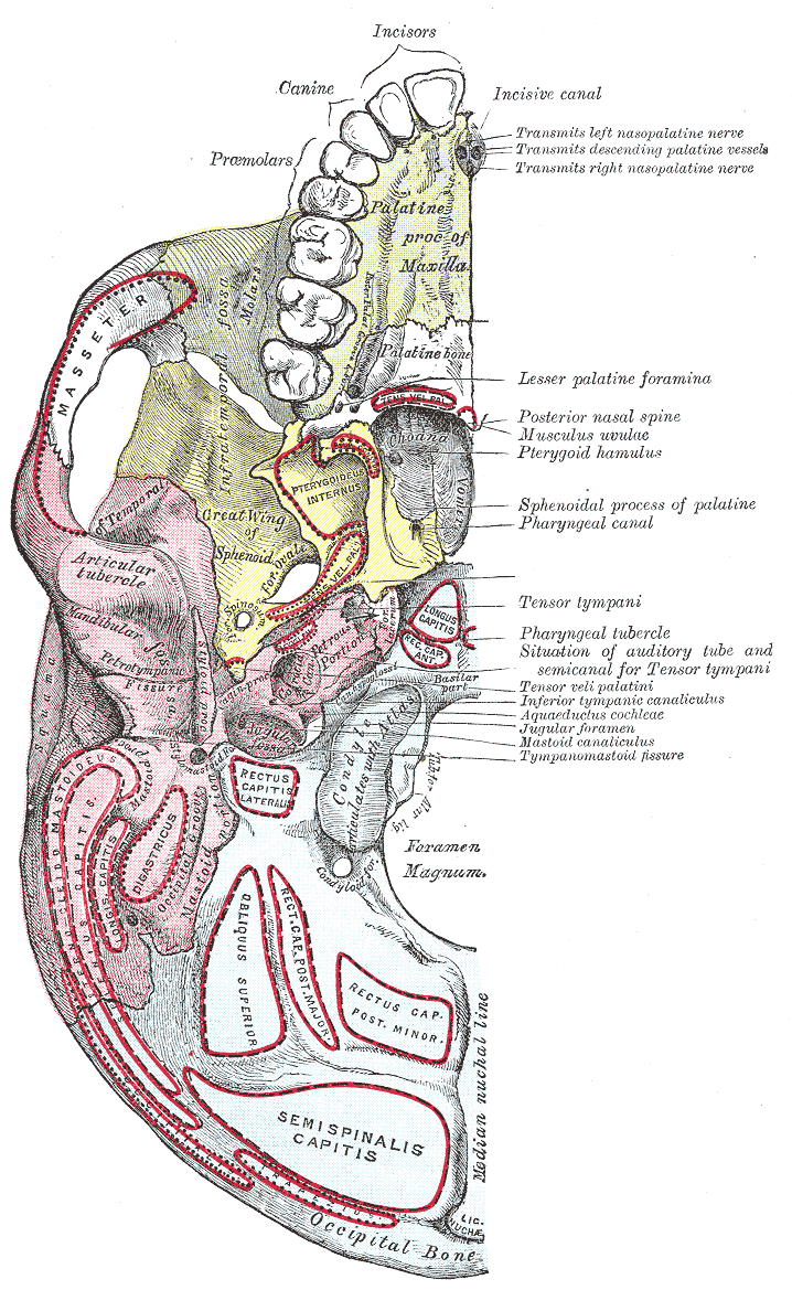
The base of skull, also known as the cranial base or the cranial floor, is the most inferior area of the skull. It is composed of the endocranium and the lower parts of the calvaria. Structure Structures found at the base of the skull are for example: Bones There are five bones that make up the base of the skull: * Ethmoid bone *Sphenoid bone *Occipital bone *Frontal bone *Temporal bone Sinuses * Occipital sinus * Superior sagittal sinus * Superior petrosal sinus Foramina of the skull * Foramen cecum * Optic foramen * Foramen lacerum * Foramen rotundum *Foramen magnum * Foramen ovale * Jugular foramen * Internal auditory meatus * Mastoid foramen * Sphenoidal emissary foramen * Foramen spinosum Sutures * Frontoethmoidal suture * Sphenofrontal suture * Sphenopetrosal suture *Sphenoethmoidal suture * Petrosquamous suture *Sphenosquamosal suture Other * Sphenoidal lingula * Subarcuate fossa *Dorsum sellae * Jugular process * Petro-occipital fissure * Condylar canal * Jug ... [...More Info...] [...Related Items...] OR: [Wikipedia] [Google] [Baidu] |
Sphenoid Bone
The sphenoid bone is an unpaired bone of the neurocranium. It is situated in the middle of the skull towards the front, in front of the basilar part of the occipital bone. The sphenoid bone is one of the seven bones that articulate to form the orbit. Its shape somewhat resembles that of a butterfly or bat with its wings extended. Structure It is divided into the following parts: * a median portion, known as the body of sphenoid bone, containing the sella turcica, which houses the pituitary gland as well as the paired paranasal sinuses, the sphenoidal sinuses * two greater wings on the lateral side of the body and two lesser wings from the anterior side. * Pterygoid processes of the sphenoides, directed downwards from the junction of the body and the greater wings. Two sphenoidal conchae are situated at the anterior and inferior part of the body. Intrinsic ligaments of the sphenoid The more important of these are: * the pterygospinous, stretching between the spina angu ... [...More Info...] [...Related Items...] OR: [Wikipedia] [Google] [Baidu] |
Lateral Fissure
In neuroanatomy, the lateral sulcus (also called Sylvian fissure, after Franciscus Sylvius, or lateral fissure) is one of the most prominent features of the human brain. The lateral sulcus is a deep fissure in each hemisphere that separates the frontal and parietal lobes from the temporal lobe. The insular cortex lies deep within the lateral sulcus. Anatomy The lateral sulcus divides both the frontal lobe and parietal lobe above from the temporal lobe below. It is in both hemispheres of the brain. The lateral sulcus is one of the earliest-developing sulci of the human brain. It first appears around the fourteenth gestational week. The insular cortex lies deep within the lateral sulcus. The lateral sulcus has a number of side branches. Two of the most prominent and most regularly found are the ascending (also called vertical) ramus and the horizontal ramus of the lateral fissure, which subdivide the inferior frontal gyrus. The lateral sulcus also contains the transverse t ... [...More Info...] [...Related Items...] OR: [Wikipedia] [Google] [Baidu] |
Brain
A brain is an organ that serves as the center of the nervous system in all vertebrate and most invertebrate animals. It is located in the head, usually close to the sensory organs for senses such as vision. It is the most complex organ in a vertebrate's body. In a human, the cerebral cortex contains approximately 14–16 billion neurons, and the estimated number of neurons in the cerebellum is 55–70 billion. Each neuron is connected by synapses to several thousand other neurons. These neurons typically communicate with one another by means of long fibers called axons, which carry trains of signal pulses called action potentials to distant parts of the brain or body targeting specific recipient cells. Physiologically, brains exert centralized control over a body's other organs. They act on the rest of the body both by generating patterns of muscle activity and by driving the secretion of chemicals called hormones. This centralized control allows rapid and coordinated responses ... [...More Info...] [...Related Items...] OR: [Wikipedia] [Google] [Baidu] |
Tentorium Cerebelli
The cerebellar tentorium or tentorium cerebelli (Latin for "tent of the cerebellum") is an extension of the dura mater that separates the cerebellum from the inferior portion of the occipital lobes. Structure The cerebellar tentorium is an arched lamina, elevated in the middle, and inclining downward toward the circumference. It covers the top of the cerebellum, and supports the occipital lobes of the brain. Its anterior border is free and concave, and bounds a large oval opening, the tentorial incisure, through which pass the cerebral peduncles. It is attached, behind, by its convex border, to the transverse ridges upon the inner surface of the occipital bone, and there encloses the transverse sinuses; in front, to the superior angle of the petrous part of the temporal bone on either side, enclosing the superior petrosal sinuses. At the apex of the petrous part of the temporal bone the free and attached borders meet, and, crossing one another, are continued forward to ... [...More Info...] [...Related Items...] OR: [Wikipedia] [Google] [Baidu] |
Middle Clinoid Process
The anterior boundary of the sella turcica is completed by two small eminences, one on either side, called the middle clinoid processes. It is found lateral to the sella turcica. Etymology Clinoid likely comes from the Greek root ''klinein'' or the Latin Latin (, or , ) is a classical language belonging to the Italic branch of the Indo-European languages. Latin was originally a dialect spoken in the lower Tiber area (then known as Latium) around present-day Rome, but through the power of the ... ''clinare'', both meaning "sloped" as in "inclined." References Bones of the head and neck {{musculoskeletal-stub ... [...More Info...] [...Related Items...] OR: [Wikipedia] [Google] [Baidu] |
Internal Carotid Artery
The internal carotid artery (Latin: arteria carotis interna) is an artery in the neck which supplies the anterior circulation of the brain. In human anatomy, the internal and external carotids arise from the common carotid arteries, where these bifurcate at cervical vertebrae C3 or C4. The internal carotid artery supplies the brain, including the eyes, while the external carotid nourishes other portions of the head, such as the face, scalp, skull, and meninges. Classification Terminologia Anatomica in 1998 subdivided the artery into four parts: "cervical", "petrous", "cavernous", and "cerebral". However, in clinical settings, the classification system of the internal carotid artery usually follows the 1996 recommendations by Bouthillier, describing seven anatomical segments of the internal carotid artery, each with a corresponding alphanumeric identifier—C1 cervical, C2 petrous, C3 lacerum, C4 cavernous, C5 clinoid, C6 ophthalmic, and C7 communicating. The Bouthillier nome ... [...More Info...] [...Related Items...] OR: [Wikipedia] [Google] [Baidu] |
Foramen
In anatomy and osteology, a foramen (; in Merriam-Webster Online Dictionary '. plural foramina, or foramens ) is an open hole that is present in extant or extinct amniotes. Foramina inside the body of typically allow , |
Dura Mater
In neuroanatomy, dura mater is a thick membrane made of dense irregular connective tissue that surrounds the brain and spinal cord. It is the outermost of the three layers of membrane called the meninges that protect the central nervous system. The other two meningeal layers are the arachnoid mater and the pia mater. It envelops the arachnoid mater, which is responsible for keeping in the cerebrospinal fluid. It is derived primarily from the neural crest cell population, with postnatal contributions of the paraxial mesoderm. Structure The dura mater has several functions and layers. The dura mater is a membrane that envelops the arachnoid mater. It surrounds and supports the dural sinuses (also called dural venous sinuses, cerebral sinuses, or cranial sinuses) and carries blood from the brain toward the heart. Cranial dura mater has two layers called '' lamellae'', a superficial layer (also called the periosteal layer), which serves as the skull's inner periosteum, called t ... [...More Info...] [...Related Items...] OR: [Wikipedia] [Google] [Baidu] |
Posterior Clinoid Process
In the sphenoid bone, the anterior boundary of the sella turcica is completed by two small eminences, one on either side, called the anterior clinoid processes, while the posterior boundary is formed by a square-shaped plate of bone, the dorsum sellæ, ending at its superior angles in two tubercles, the posterior clinoid processes, the size and form of which vary considerably in different individuals. The posterior clinoid processes deepen the sella turcica, and give attachment to the tentorium cerebelli. The petroclinoid ligament The petroclinoid ligament is a fold of dura matter. It extends between the posterior clinoid process and anterior clinoid process and the petrosal part of the temporal bone The temporal bones are situated at the sides and base of the skull, and lateral to the temporal lobes of the cerebral cortex. The temporal bones are overlaid by the sides of the head known as the temples, and house the structures of the ears. ... of the skull. There are two s ... [...More Info...] [...Related Items...] OR: [Wikipedia] [Google] [Baidu] |
Temporal Bone
The temporal bones are situated at the sides and base of the skull, and lateral to the temporal lobes of the cerebral cortex. The temporal bones are overlaid by the sides of the head known as the temples, and house the structures of the ears. The lower seven cranial nerves and the major vessels to and from the brain traverse the temporal bone. Structure The temporal bone consists of four parts— the squamous, mastoid, petrous and tympanic parts. The squamous part is the largest and most superiorly positioned relative to the rest of the bone. The zygomatic process is a long, arched process projecting from the lower region of the squamous part and it articulates with the zygomatic bone. Posteroinferior to the squamous is the mastoid part. Fused with the squamous and mastoid parts and between the sphenoid and occipital bones lies the petrous part, which is shaped like a pyramid. The tympanic part is relatively small and lies inferior to the squamous part, anterior to t ... [...More Info...] [...Related Items...] OR: [Wikipedia] [Google] [Baidu] |


