|
Anococcygeal Nerve
The anococcygeal nerve is a nerve in the pelvis which provides sensory innervation to the skin over the coccyx. The number of anococcygeal nerves vary between one and three. Structure The anococcygeal nerve arises from the coccygeal plexus. It pierces the sacrotuberous ligament to supply the skin in the region of the coccyx as well as the sacrococcygeal joint. See also * Coccyx * Coccydynia (coccyx pain, tailbone pain) * Ganglion impar * Sacral plexus In human anatomy, the sacral plexus is a nerve plexus which provides motor and sensory nerves for the posterior thigh, most of the lower leg and foot, and part of the pelvis. It is part of the lumbosacral plexus and emerges from the lumbar ver ... References External linksCoccyx pain, tailbone pain, coccydynia(Peer-reviewed medical chapter, available free online at eMedicine) Nerves of the lower limb and lower torso {{Neuroanatomy-stub ... [...More Info...] [...Related Items...] OR: [Wikipedia] [Google] [Baidu] |
Sacral Plexus
In human anatomy, the sacral plexus is a nerve plexus which provides motor and sensory nerves for the posterior thigh, most of the lower leg and foot, and part of the pelvis. It is part of the lumbosacral plexus and emerges from the lumbar vertebrae and sacral vertebrae (L4-S4).''Thieme Atlas of Anatomy'' (2006), pp 470-471 A sacral plexopathy is a disorder affecting the nerves of the sacral plexus, usually caused by trauma, nerve compression, vascular disease, or infection. Symptoms may include pain, loss of motor control, and sensory deficits. Structure The sacral plexus is formed by: * the lumbosacral trunk * the anterior division of the first sacral nerve * portions of the anterior divisions of the second and third sacral nerves The nerves forming the sacral plexus converge toward the lower part of the greater sciatic foramen, and unite to form a flattened band, from the anterior and posterior surfaces of which several branches arise. The band itself is continued as th ... [...More Info...] [...Related Items...] OR: [Wikipedia] [Google] [Baidu] |
Pudendal Plexus (nerves)
The pudendal plexus is a term used for a compound structure consisting of sacral spinal nerves. Some sources describe it as S2-S4 of the lumbosacral plexus The anterior divisions of the lumbar nerves, sacral nerves, and coccygeal nerve form the lumbosacral plexus, the first lumbar nerve being frequently joined by a branch from the twelfth thoracic. For descriptive purposes this plexus is usually d .... In some older texts, it is a rough approximation of the coccygeal plexus. References Nerve plexus {{neuroanatomy-stub ... [...More Info...] [...Related Items...] OR: [Wikipedia] [Google] [Baidu] |
Coccygeal Plexus
The coccygeal plexus is a nerve plexus near the coccyx bone. Structure This plexus is formed by the ventral rami of the fourth and fifth sacral nerves, S4 & S5, and the ventral ramus of the coccygeal nerve, Co. The relative contributions of S4 and S5 are minor and major, respectively. It gives rise to the anococcygeal nerve. See also * Coccydynia (coccyx pain, tailbone pain) * Ganglion impar * Sacral plexus In human anatomy, the sacral plexus is a nerve plexus which provides motor and sensory nerves for the posterior thigh, most of the lower leg and foot, and part of the pelvis. It is part of the lumbosacral plexus and emerges from the lumbar ver ... References External linksDescription at uams.edu (Peer-reviewed medical chapter, available free online at eMedicine) Nerve plex ... [...More Info...] [...Related Items...] OR: [Wikipedia] [Google] [Baidu] |
Nerve
A nerve is an enclosed, cable-like bundle of nerve fibers (called axons) in the peripheral nervous system. A nerve transmits electrical impulses. It is the basic unit of the peripheral nervous system. A nerve provides a common pathway for the electrochemical nerve impulses called action potentials that are transmitted along each of the axons to peripheral organs or, in the case of sensory nerves, from the periphery back to the central nervous system. Each axon, within the nerve, is an extension of an individual neuron, along with other supportive cells such as some Schwann cells that coat the axons in myelin. Within a nerve, each axon is surrounded by a layer of connective tissue called the endoneurium. The axons are bundled together into groups called fascicles, and each fascicle is wrapped in a layer of connective tissue called the perineurium. Finally, the entire nerve is wrapped in a layer of connective tissue called the epineurium. Nerve cells (often called neurons ... [...More Info...] [...Related Items...] OR: [Wikipedia] [Google] [Baidu] |
Pelvis
The pelvis (plural pelves or pelvises) is the lower part of the trunk, between the abdomen and the thighs (sometimes also called pelvic region), together with its embedded skeleton (sometimes also called bony pelvis, or pelvic skeleton). The pelvic region of the trunk includes the bony pelvis, the pelvic cavity (the space enclosed by the bony pelvis), the pelvic floor, below the pelvic cavity, and the perineum, below the pelvic floor. The pelvic skeleton is formed in the area of the back, by the sacrum and the coccyx and anteriorly and to the left and right sides, by a pair of hip bones. The two hip bones connect the spine with the lower limbs. They are attached to the sacrum posteriorly, connected to each other anteriorly, and joined with the two femurs at the hip joints. The gap enclosed by the bony pelvis, called the pelvic cavity, is the section of the body underneath the abdomen and mainly consists of the reproductive organs (sex organs) and the rectum, while the p ... [...More Info...] [...Related Items...] OR: [Wikipedia] [Google] [Baidu] |
Coccyx
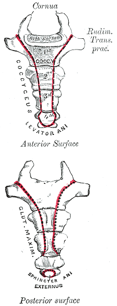
The coccyx ( : coccyges or coccyxes), commonly referred to as the tailbone, is the final segment of the vertebral column in all apes, and analogous structures in certain other mammals such as horses. In tailless primates (e.g. humans and other great apes) since '' Nacholapithecus'' (a Miocene hominoid),Nakatsukasa 2004, ''Acquisition of bipedalism'' (SeFig. 5entitled ''First coccygeal/caudal vertebra in short-tailed or tailless primates.''.) the coccyx is the remnant of a vestigial tail. In animals with bony tails, it is known as ''tailhead'' or ''dock'', in bird anatomy as ''tailfan''. It comprises three to five separate or fused coccygeal vertebrae below the sacrum, attached to the sacrum by a fibrocartilaginous joint, the sacrococcygeal symphysis, which permits limited movement between the sacrum and the coccyx. Structure The coccyx is formed of three, four or five rudimentary vertebrae. It articulates superiorly with the sacrum. In each of the first three segm ... [...More Info...] [...Related Items...] OR: [Wikipedia] [Google] [Baidu] |
Coccygeal Plexus
The coccygeal plexus is a nerve plexus near the coccyx bone. Structure This plexus is formed by the ventral rami of the fourth and fifth sacral nerves, S4 & S5, and the ventral ramus of the coccygeal nerve, Co. The relative contributions of S4 and S5 are minor and major, respectively. It gives rise to the anococcygeal nerve. See also * Coccydynia (coccyx pain, tailbone pain) * Ganglion impar * Sacral plexus In human anatomy, the sacral plexus is a nerve plexus which provides motor and sensory nerves for the posterior thigh, most of the lower leg and foot, and part of the pelvis. It is part of the lumbosacral plexus and emerges from the lumbar ver ... References External linksDescription at uams.edu (Peer-reviewed medical chapter, available free online at eMedicine) Nerve plex ... [...More Info...] [...Related Items...] OR: [Wikipedia] [Google] [Baidu] |
Sacrotuberous Ligament
The sacrotuberous ligament (great or posterior sacrosciatic ligament) is situated at the lower and back part of the pelvis. It is flat, and triangular in form; narrower in the middle than at the ends. Structure It runs from the sacrum (the lower transverse sacral tubercles, the inferior margins sacrum and the upper coccyx) to the tuberosity of the ischium. It is a remnant of part of Biceps femoris muscle. The sacrotuberous ligament is attached by its broad base to the posterior superior iliac spine, the posterior sacroiliac ligaments (with which it is partly blended), to the lower transverse sacral tubercles and the lateral margins of the lower sacrum and upper coccyx. Its oblique fibres descend laterally, converging to form a thick, narrow band that widens again below and is attached to the medial margin of the ischial tuberosity. It then spreads along the ischial ramus as the falciform process, whose concave edge blends with the fascial sheath of the internal pudendal vessels ... [...More Info...] [...Related Items...] OR: [Wikipedia] [Google] [Baidu] |
Sacrococcygeal Joint
The sacrococcygeal symphysis (sacrococcygeal articulation, articulation of the sacrum and coccyx) is an amphiarthrodial joint, formed between the oval surface at the apex of the sacrum, and the base of the coccyx. It is a slightly moveable jointMorris (2005), p 59 which is frequently, partially or completely, obliterated in old age,Palastanga (2006), p 334 homologous with the joints between the bodies of the vertebrae. Disc The sacrococcygeal disc or interosseus ligamentHuijbregts (2001), p 13 is similar to the intervertebral discs but thinner, thicker in front and behind than at the sides, and with a firmer texture. The articular surfaces are elliptical with longer transversal axes. The surface on the sacrum is convex and that on the coccyx concave. Occasionally the coccyx is freely movable on the sacrum, most notably during pregnancy; in such cases a synovial membrane is present. Ligaments The joint is strengthened by a series of ligaments: * The ventral or anterior sacro ... [...More Info...] [...Related Items...] OR: [Wikipedia] [Google] [Baidu] |
Coccyx
The coccyx ( : coccyges or coccyxes), commonly referred to as the tailbone, is the final segment of the vertebral column in all apes, and analogous structures in certain other mammals such as horses. In tailless primates (e.g. humans and other great apes) since '' Nacholapithecus'' (a Miocene hominoid),Nakatsukasa 2004, ''Acquisition of bipedalism'' (SeFig. 5entitled ''First coccygeal/caudal vertebra in short-tailed or tailless primates.''.) the coccyx is the remnant of a vestigial tail. In animals with bony tails, it is known as ''tailhead'' or ''dock'', in bird anatomy as ''tailfan''. It comprises three to five separate or fused coccygeal vertebrae below the sacrum, attached to the sacrum by a fibrocartilaginous joint, the sacrococcygeal symphysis, which permits limited movement between the sacrum and the coccyx. Structure The coccyx is formed of three, four or five rudimentary vertebrae. It articulates superiorly with the sacrum. In each of the first three segm ... [...More Info...] [...Related Items...] OR: [Wikipedia] [Google] [Baidu] |
Coccydynia
Coccydynia is a medical term meaning pain in the coccyx or tailbone area, often brought on by a fall onto the coccyx or by persistent irritation usually from sitting. Synonyms Coccydynia is also known as coccygodynia, coccygeal pain, coccyx pain, or coccalgia. Anatomy Structure Coccydynia occurs in the lowest part of the spine, the coccyx, which is believed to be a vestigial tail, or in other words the “tail bone”. The name coccyx is derived from the Greek word for cuckoo due to its beak like appearance. The coccyx itself is made up of 3 to 5 vertebrae, some of which may be fused together. The ventral side of the coccyx is slightly concave whereas the dorsal aspect is slightly convex. Both of these sides have transverse grooves that show where the vestigial coccygeal units had previously fused. The coccyx attaches to the sacrum from the dorsal grooves, with the attachment being either a symphysis or as a true synovial joint, and also to the gluteus maximus muscle, the cocc ... [...More Info...] [...Related Items...] OR: [Wikipedia] [Google] [Baidu] |
Ganglion Impar
The pelvic portion of each sympathetic trunk is situated in front of the sacrum The sacrum (plural: ''sacra'' or ''sacrums''), in human anatomy, is a large, triangular bone at the base of the spine that forms by the fusing of the sacral vertebrae (S1S5) between ages 18 and 30. The sacrum situates at the upper, back part ..., medial to the anterior sacral foramina. It consists of four or five small sacral ganglia, connected together by interganglionic cords, and continuous above with the abdominal portion. Below, the two pelvic sympathetic trunks converge, and end on the front of the coccyx in a small ganglion, the ganglion impar, also known as azygos or ganglion of Walther. Clinical significance A study found that in some cases a single injection of nerve block at the ganglion impar offered complete relief from coccydynia. References * Munir MA, Zhang J, Ahmad M. (2004)A modified needle-inside-needle technique for the ganglion impar block. Can J Anaesth. 2004 N ... [...More Info...] [...Related Items...] OR: [Wikipedia] [Google] [Baidu] |


