|
Annular Ligament Of Radius
The annular ligament (orbicular ligament) is a strong band of fibers that encircles the head of the radius, and retains it in contact with the radial notch of the ulna.'' Gray's Anatomy'' (1918), see infobox Per '' Terminologia Anatomica 1998'', the spelling is "anular", but the spelling "annular" is frequently encountered. Indeed, the most recent version of ''Terminologia Anatomica'' (2019) uses "annular" as the preferred English spelling. Anatomy The annular ligament is attached by both its ends to the anterior and posterior margins of the radial notch of the ulna, together with which it forms the articular surface that surrounds the head and neck of the radius. The ligament is strong and well defined, yet its flexibility permits the slightly oval head of the radius to rotate freely during pronation and supination. The head of the radius is wider than the bone's neck, and, because the annular ligament embraces both, the radial head is "trapped" inside the ligament which thus ac ... [...More Info...] [...Related Items...] OR: [Wikipedia] [Google] [Baidu] |
Radius (bone)
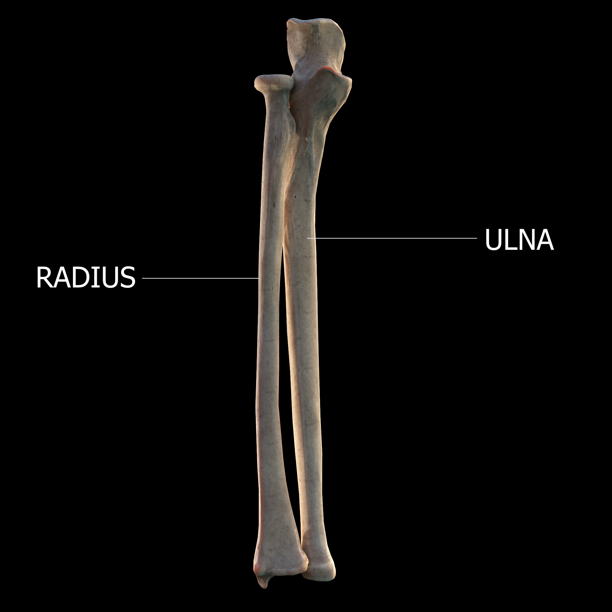
The radius or radial bone is one of the two large bones of the forearm, the other being the ulna. It extends from the lateral side of the elbow to the thumb side of the wrist and runs parallel to the ulna. The ulna is usually slightly longer than the radius, but the radius is thicker. Therefore the radius is considered to be the larger of the two. It is a long bone, prism-shaped and slightly curved longitudinally. The radius is part of two joints: the elbow and the wrist. At the elbow, it joins with the capitulum of the humerus, and in a separate region, with the ulna at the radial notch. At the wrist, the radius forms a joint with the ulna bone. The corresponding bone in the lower leg is the fibula. Structure The long narrow medullary cavity is enclosed in a strong wall of compact bone. It is thickest along the interosseous border and thinnest at the extremities, same over the cup-shaped articular surface (fovea) of the head. The trabeculae of the spongy t ... [...More Info...] [...Related Items...] OR: [Wikipedia] [Google] [Baidu] |
Head Of Radius
The head of the radius has a cylindrical form, and on its upper surface is a shallow cup or fovea for articulation with the capitulum of the humerus. The circumference of the head is smooth; it is broad medially where it articulates with the radial notch of the ulna, narrow in the rest of its extent, which is embraced by the annular ligament.'' Gray's Anatomy'' (1918), see infobox Articular surfaces The head of the radius is shaped to articulate with a complex of articular surfaces during both flexion-extension at the elbow and supination-pronation in the forearm: Humeroradial joint The head's proximal surface is concave and cup-shaped to correspond to the spherical surface of the capitulum of the humerus. The radius can thus glide on the capitulum during elbow flexion-extension while simultaneously rotate about its own main axis during supination-pronation. Between the capitulum and the trochlea of the humerus is the capitulotrochlear groove. A semi-lunar surface around t ... [...More Info...] [...Related Items...] OR: [Wikipedia] [Google] [Baidu] |
Epiphyseal Plate
The epiphyseal plate (or epiphysial plate, physis, or growth plate) is a hyaline cartilage plate in the metaphysis at each end of a long bone. It is the part of a long bone where new bone growth takes place; that is, the whole bone is alive, with maintenance remodeling throughout its existing bone tissue, but the growth plate is the place where the long bone grows longer (adds length). The plate is only found in children and adolescents; in adults, who have stopped growing, the plate is replaced by an epiphyseal line. This replacement is known as epiphyseal closure or growth plate fusion. Complete fusion can occur as early as 12 for girls (with the most common being 14-15 years for girls) and as early as 14 for boys (with the most common being 15–17 years for boys). Structure Development Endochondral ossification is responsible for the initial bone development from cartilage in utero and infants and the longitudinal growth of long bones in the epiphyseal plate. The plate' ... [...More Info...] [...Related Items...] OR: [Wikipedia] [Google] [Baidu] |
Quadrate Ligament
In human anatomy, the quadrate ligament or ligament of Denucé is one of the ligaments of the proximal radioulnar joint in the upper forearm. Structure The quadrate ligament is a fibrous band attached to the inferior border of the radial notch on the ulna and to the neck of the radius. Its borders are strengthened by fibers from the upper border of the annular ligament. The ligament is long, wide, and thick. Function The quadrate ligament reinforces the inferior part of the capsule of the elbow joint and contributes to joint stability by securing the proximal radius against the radial notch and by restricting excessive supination (10–20° restriction) and, to a lesser degree, pronation Motion, the process of movement, is described using specific anatomical terms. Motion includes movement of organs, joints, limbs, and specific sections of the body. The terminology used describes this motion according to its direction relativ ... (5–8°). History The quadrate ligame ... [...More Info...] [...Related Items...] OR: [Wikipedia] [Google] [Baidu] |
Hyaline Cartilage
Hyaline cartilage is the glass-like (hyaline) and translucent cartilage found on many joint surfaces. It is also most commonly found in the ribs, nose, larynx, and trachea. Hyaline cartilage is pearl-gray in color, with a firm consistency and has a considerable amount of collagen. It contains no nerves or blood vessels, and its structure is relatively simple. Structure Hyaline cartilage is covered externally by a fibrous membrane known as the perichondrium or, when it's along articulating surfaces, the synovial membrane. This membrane contains vessels that provide the cartilage with nutrition through diffusion. Hyaline cartilage matrix is primarily made of type II collagen and chondroitin sulphate, both of which are also found in elastic cartilage. Hyaline cartilage exists on the sternal ends of the ribs, in the larynx, trachea, and bronchi, and on the articulating surfaces of bones. It gives the structures a definite but pliable form. The presence of collagen fibres makes ... [...More Info...] [...Related Items...] OR: [Wikipedia] [Google] [Baidu] |
Fibrocartilage
Fibrocartilage consists of a mixture of white fibrous tissue and cartilaginous tissue in various proportions. It owes its inflexibility and toughness to the former of these constituents, and its elasticity to the latter. It is the only type of cartilage that contains type I collagen in addition to the normal type II. Structure The extracellular matrix of fibrocartilage is mainly made from type I collagen secreted by chondroblasts. Locations of fibrocartilage in the human body * secondary cartilaginous joints: ** pubic symphysis ** annulus fibrosis of intervertebral discs ** manubriosternal joint * glenoid labrum of shoulder joint * acetabular labrum of hip joint * medial and lateral menisci of the knee joint * location where tendons and ligaments A ligament is the fibrous connective tissue that connects bones to other bones. It is also known as ''articular ligament'', ''articular larua'', ''fibrous ligament'', or ''true ligament''. Other ligaments in the bo ... [...More Info...] [...Related Items...] OR: [Wikipedia] [Google] [Baidu] |
Radial Collateral Ligament Of Elbow Joint
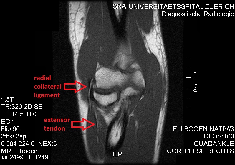
The radial collateral ligament (RCL), lateral collateral ligament (LCL), or external lateral ligamentAs opposed to the "internal lateral ligament", better known as the medial or ulnar collateral ligament is a ligament in the elbow on the side of the radius. Structure The composition of the triangular ligamentous structure on the lateral side of the elbow varies widely between individuals, see alsFigure 4/ref> and can be considered either a single ligament, in which case multiple distal attachments are generally mentioned and the annular ligament is described separately, or as several separate ligaments, in which case parts of those ligaments are often described as indistinguishable from each other. In the latter case, the ligaments are collectively referred to as the lateral collateral ligament complex (LCLC), consisting of four ligaments: * the radial collateral ligament roper(RCL), from the lateral epicondyle to the annular ligament deep to the common extensor tendon * the ... [...More Info...] [...Related Items...] OR: [Wikipedia] [Google] [Baidu] |
Forearm
The forearm is the region of the upper limb between the elbow and the wrist. The term forearm is used in anatomy to distinguish it from the arm, a word which is most often used to describe the entire appendage of the upper limb, but which in anatomy, technically, means only the region of the upper arm, whereas the lower "arm" is called the forearm. It is homologous to the region of the leg that lies between the knee and the ankle joints, the crus. The forearm contains two long bones, the radius and the ulna, forming the two radioulnar joints. The interosseous membrane connects these bones. Ultimately, the forearm is covered by skin, the anterior surface usually being less hairy than the posterior surface. The forearm contains many muscles, including the flexors and extensors of the wrist, flexors and extensors of the digits, a flexor of the elbow ( brachioradialis), and pronators and supinators that turn the hand to face down or upwards, respectively. In cross-section, ... [...More Info...] [...Related Items...] OR: [Wikipedia] [Google] [Baidu] |
Supination
Motion, the process of movement, is described using specific anatomical terms. Motion includes movement of organs, joints, limbs, and specific sections of the body. The terminology used describes this motion according to its direction relative to the anatomical position of the body parts involved. Anatomists and others use a unified set of terms to describe most of the movements, although other, more specialized terms are necessary for describing unique movements such as those of the hands, feet, and eyes. In general, motion is classified according to the anatomical plane it occurs in. ''Flexion'' and ''extension'' are examples of ''angular'' motions, in which two axes of a joint are brought closer together or moved further apart. ''Rotational'' motion may occur at other joints, for example the shoulder, and are described as ''internal'' or ''external''. Other terms, such as ''elevation'' and ''depression'', describe movement above or below the horizontal plane. Many anatomi ... [...More Info...] [...Related Items...] OR: [Wikipedia] [Google] [Baidu] |
Radius (bone)
The radius or radial bone is one of the two large bones of the forearm, the other being the ulna. It extends from the lateral side of the elbow to the thumb side of the wrist and runs parallel to the ulna. The ulna is usually slightly longer than the radius, but the radius is thicker. Therefore the radius is considered to be the larger of the two. It is a long bone, prism-shaped and slightly curved longitudinally. The radius is part of two joints: the elbow and the wrist. At the elbow, it joins with the capitulum of the humerus, and in a separate region, with the ulna at the radial notch. At the wrist, the radius forms a joint with the ulna bone. The corresponding bone in the lower leg is the fibula. Structure The long narrow medullary cavity is enclosed in a strong wall of compact bone. It is thickest along the interosseous border and thinnest at the extremities, same over the cup-shaped articular surface (fovea) of the head. The trabeculae of the spongy t ... [...More Info...] [...Related Items...] OR: [Wikipedia] [Google] [Baidu] |
Pronation
Motion, the process of movement, is described using specific anatomical terms. Motion includes movement of organs, joints, limbs, and specific sections of the body. The terminology used describes this motion according to its direction relative to the anatomical position of the body parts involved. Anatomists and others use a unified set of terms to describe most of the movements, although other, more specialized terms are necessary for describing unique movements such as those of the hands, feet, and eyes. In general, motion is classified according to the anatomical plane it occurs in. ''Flexion'' and ''extension'' are examples of ''angular'' motions, in which two axes of a joint are brought closer together or moved further apart. ''Rotational'' motion may occur at other joints, for example the shoulder, and are described as ''internal'' or ''external''. Other terms, such as ''elevation'' and ''depression'', describe movement above or below the horizontal plane. Many anatomi ... [...More Info...] [...Related Items...] OR: [Wikipedia] [Google] [Baidu] |
Joint
A joint or articulation (or articular surface) is the connection made between bones, ossicles, or other hard structures in the body which link an animal's skeletal system into a functional whole.Saladin, Ken. Anatomy & Physiology. 7th ed. McGraw-Hill Connect. Webp.274/ref> They are constructed to allow for different degrees and types of movement. Some joints, such as the knee, elbow, and shoulder, are self-lubricating, almost frictionless, and are able to withstand compression and maintain heavy loads while still executing smooth and precise movements. Other joints such as sutures between the bones of the skull permit very little movement (only during birth) in order to protect the brain and the sense organs. The connection between a tooth and the jawbone is also called a joint, and is described as a fibrous joint known as a gomphosis. Joints are classified both structurally and functionally. Classification The number of joints depends on if sesamoids are included, age of ... [...More Info...] [...Related Items...] OR: [Wikipedia] [Google] [Baidu] |


