|
Analyze (imaging Software)
Analyze is a software package developed by the Biomedical Imaging Resource (BIR) at Mayo Clinic for multi-dimensional display, processing, and measurement of multi-modality biomedical images. It is a commercial program and is used for medical tomographic scans from magnetic resonance imaging, computed tomography and positron emission tomography. The Analyze 7.5 file format has been widely used in the functional neuroimaging field, and other programs such as SPM, FreeSurfer, AIR, MRIcro and Mango are able to read and write the format. The files can be used to store voxel-based volumes. One data item consists of two files: One file with the actual data in a binary format with the filename extension A filename extension, file name extension or file extension is a suffix to the name of a computer file (e.g., .txt, .docx, .md). The extension indicates a characteristic of the file contents or its intended use. A filename extension is typically d ... .img and another file (''header'' ... [...More Info...] [...Related Items...] OR: [Wikipedia] [Google] [Baidu] |
Biomedical Imaging Resource
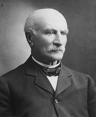
The Mayo Clinic () is a nonprofit American academic medical center focused on integrated health care, education, and research. It employs over 4,500 physicians and scientists, along with another 58,400 administrative and allied health staff, across three major campuses: Rochester, Minnesota; Jacksonville, Florida; and Phoenix/Scottsdale, Arizona. The practice specializes in treating difficult cases through tertiary care and destination medicine. It is home to the top-15 ranked Mayo Clinic Alix School of Medicine in addition to many of the highest regarded residency education programs in the United States. It spends over $660 million a year on research and has more than 3,000 full-time research personnel. William Worrall Mayo settled his family in Rochester in 1864 and opened a sole proprietorship medical practice that evolved under his sons, Will and Charlie Mayo, along with practice partners Stinchfield, Graham, Plummer, Millet, Judd, and Balfour, into Mayo Clinic. Today ... [...More Info...] [...Related Items...] OR: [Wikipedia] [Google] [Baidu] |
Mayo Clinic
The Mayo Clinic () is a nonprofit American academic medical center focused on integrated health care, education, and research. It employs over 4,500 physicians and scientists, along with another 58,400 administrative and allied health staff, across three major campuses: Rochester, Minnesota; Jacksonville, Florida; and Phoenix/Scottsdale, Arizona. The practice specializes in treating difficult cases through tertiary care and destination medicine. It is home to the top-15 ranked Mayo Clinic Alix School of Medicine in addition to many of the highest regarded residency education programs in the United States. It spends over $660 million a year on research and has more than 3,000 full-time research personnel. William Worrall Mayo settled his family in Rochester in 1864 and opened a sole proprietorship medical practice that evolved under his sons, Will and Charlie Mayo, along with practice partners Stinchfield, Graham, Plummer, Millet, Judd, and Balfour, into Mayo Clinic. Toda ... [...More Info...] [...Related Items...] OR: [Wikipedia] [Google] [Baidu] |
Magnetic Resonance Imaging
Magnetic resonance imaging (MRI) is a medical imaging technique used in radiology to form pictures of the anatomy and the physiological processes of the body. MRI scanners use strong magnetic fields, magnetic field gradients, and radio waves to generate images of the organs in the body. MRI does not involve X-rays or the use of ionizing radiation, which distinguishes it from CT and PET scans. MRI is a medical application of nuclear magnetic resonance (NMR) which can also be used for imaging in other NMR applications, such as NMR spectroscopy. MRI is widely used in hospitals and clinics for medical diagnosis, staging and follow-up of disease. Compared to CT, MRI provides better contrast in images of soft-tissues, e.g. in the brain or abdomen. However, it may be perceived as less comfortable by patients, due to the usually longer and louder measurements with the subject in a long, confining tube, though "Open" MRI designs mostly relieve this. Additionally, implants and oth ... [...More Info...] [...Related Items...] OR: [Wikipedia] [Google] [Baidu] |
Computed Tomography
A computed tomography scan (CT scan; formerly called computed axial tomography scan or CAT scan) is a medical imaging technique used to obtain detailed internal images of the body. The personnel that perform CT scans are called radiographers or radiology technologists. CT scanners use a rotating X-ray tube and a row of detectors placed in a gantry to measure X-ray attenuations by different tissues inside the body. The multiple X-ray measurements taken from different angles are then processed on a computer using tomographic reconstruction algorithms to produce tomographic (cross-sectional) images (virtual "slices") of a body. CT scans can be used in patients with metallic implants or pacemakers, for whom magnetic resonance imaging (MRI) is contraindicated. Since its development in the 1970s, CT scanning has proven to be a versatile imaging technique. While CT is most prominently used in medical diagnosis, it can also be used to form images of non-living objects. The 1979 Nob ... [...More Info...] [...Related Items...] OR: [Wikipedia] [Google] [Baidu] |
Positron Emission Tomography
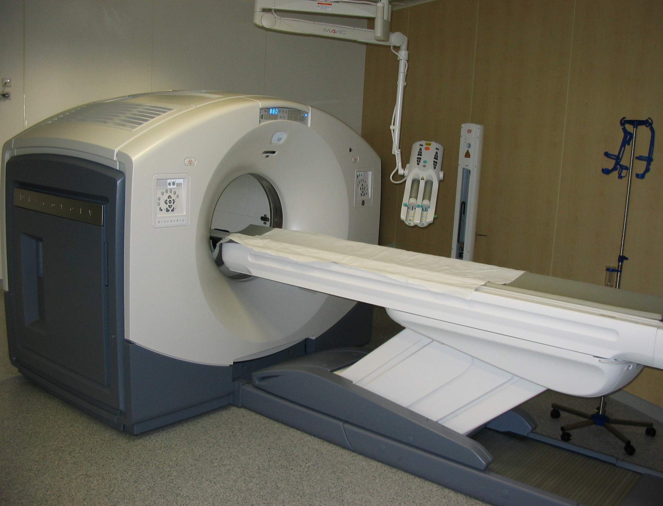
Positron emission tomography (PET) is a functional imaging technique that uses radioactive substances known as radiotracers to visualize and measure changes in Metabolism, metabolic processes, and in other physiological activities including blood flow, regional chemical composition, and absorption. Different tracers are used for various imaging purposes, depending on the target process within the body. For example, 18F-FDG, -FDG is commonly used to detect cancer, Sodium fluoride#Medical imaging, NaF is widely used for detecting bone formation, and Isotopes of oxygen#Oxygen-15, oxygen-15 is sometimes used to measure blood flow. PET is a common medical imaging, imaging technique, a Scintigraphy#Process, medical scintillography technique used in nuclear medicine. A radiopharmaceutical, radiopharmaceutical — a radioisotope attached to a drug — is injected into the body as a radioactive tracer, tracer. When the radiopharmaceutical undergoes beta plus decay, a positron is ... [...More Info...] [...Related Items...] OR: [Wikipedia] [Google] [Baidu] |
Functional Neuroimaging
Functional neuroimaging is the use of neuroimaging technology to measure an aspect of brain function, often with a view to understanding the relationship between activity in certain brain areas and specific mental functions. It is primarily used as a research tool in cognitive neuroscience, cognitive psychology, neuropsychology, and social neuroscience. Overview Common methods of functional neuroimaging include * Positron emission tomography (PET) * Functional magnetic resonance imaging (fMRI) * Electroencephalography (EEG) * Magnetoencephalography (MEG) * Functional near-infrared spectroscopy (fNIRS) * Single-photon emission computed tomography (SPECT) * Functional ultrasound imaging (fUS) PET, fMRI, fNIRS and fUS can measure localized changes in cerebral blood flow related to neural activity. These changes are referred to as ''activations''. Regions of the brain which are activated when a subject performs a particular task may play a role in the computational neuroscience, n ... [...More Info...] [...Related Items...] OR: [Wikipedia] [Google] [Baidu] |
Statistical Parametric Mapping
Statistical parametric mapping (SPM) is a statistical technique for examining differences in brain activity recorded during functional neuroimaging experiments. It was created by Karl Friston. It may alternatively refer to software created by the Wellcome Department of Imaging Neuroscience at University College London to carry out such analyses. Approach Unit of measurement Functional neuroimaging is one type of 'brain scanning'. It involves the measurement of brain activity. The measurement technique depends on the imaging technology (e.g., fMRI and PET). The scanner produces a 'map' of the area that is represented as voxels. Each voxel represents the activity of a specific volume in three-dimensional space. The exact size of a voxel varies depending on the technology. fMRI voxels typically represent a volume of 27 mm3 in an equilateral cuboid. Experimental design Researchers examine brain activity linked to a specific mental process or processes. One approach involves aski ... [...More Info...] [...Related Items...] OR: [Wikipedia] [Google] [Baidu] |
FreeSurfer
FreeSurfer is a brain imaging software package originally developed by Bruce Fischl, Anders Dale, Martin Sereno, and Doug Greve. Development and maintenance of FreeSurfer is now the primary responsibility of the Laboratory for Computational Neuroimaging at the Athinoula A. Martinos Center for Biomedical Imaging. FreeSurfer contains a set of programs with a common focus of analyzing magnetic resonance imaging (MRI) scans of brain tissue. It is an important tool in functional brain mapping and contains tools to conduct both volume based and surface based analysis. FreeSurfer includes tools for the reconstruction of topologically correct and geometrically accurate models of both the gray/white and pial surfaces, for measuring cortical thickness, surface area and folding, and for computing inter-subject registration based on the pattern of cortical folds. 57,541 copies of the FreeSurfer software package have been registered for use as of April 2022 and it is a core tool in the pr ... [...More Info...] [...Related Items...] OR: [Wikipedia] [Google] [Baidu] |
AIR (program)
The AIR (''Automated Image Registration'') is a program suite for volume-based image registration constructed by Roger P. Woods from UCLA School of Medicine. It reads and writes Analyze volume files and can work with 4x4 transformation matrices stored in its own file format with the filename extension .air. It is especially designed for neuroimaging applications and has primarily been used in research-oriented functional neuroimaging with brain scans from positron emission tomography and magnetic resonance Magnetic resonance is a process by which a physical excitation (resonance) is set up via magnetism. This process was used to develop magnetic resonance imaging and Nuclear magnetic resonance spectroscopy technology. It is also being used to ... scanners. The suite provides a number of programs for image registration with different transformation models, such as rigid-body, affine and nonlinear warping. For example, for affine transformation the registration from one b ... [...More Info...] [...Related Items...] OR: [Wikipedia] [Google] [Baidu] |
Mango (software)
Mango (Multi-Image Analysis GUI) is a non-commercial software for viewing, editing and analyzing volumetric medical images. Mango is written in Java, and distributed freely in precompiled versions for Linux, Mac OS and Microsoft Windows. It supports NIfTI, ANALYZE, NEMA and DICOM formats and is able to load and save 2D, 3D and 4D images. Mango provides tools for creation and editing of regions of interest (ROI) within the images, surface rendering, image stacking (overlaying), filtering in space domain and histogram analysis, among other functions that can be used in neuroimaging analysis for scientific (non-clinical) purposes. The software can be extended with user-defined functions ( plug-ins), which can be created using the Java language and the Mango API. See also * List of neuroimaging software Neuroimaging software is used to study the structure and function of the brain. To see an NIH Blueprint for Neuroscience Research funded clearinghouse of many of these software ... [...More Info...] [...Related Items...] OR: [Wikipedia] [Google] [Baidu] |
Voxel
In 3D computer graphics, a voxel represents a value on a regular grid in three-dimensional space. As with pixels in a 2D bitmap, voxels themselves do not typically have their position (i.e. coordinates) explicitly encoded with their values. Instead, rendering systems infer the position of a voxel based upon its position relative to other voxels (i.e., its position in the data structure that makes up a single volumetric image). In contrast to pixels and voxels, polygons are often explicitly represented by the coordinates of their vertices (as points). A direct consequence of this difference is that polygons can efficiently represent simple 3D structures with much empty or homogeneously filled space, while voxels excel at representing regularly sampled spaces that are non-homogeneously filled. Voxels are frequently used in the visualization and analysis of medical and scientific data (e.g. geographic information systems (GIS)). Some volumetric displays use voxels to describe ... [...More Info...] [...Related Items...] OR: [Wikipedia] [Google] [Baidu] |


