|
Coccyx
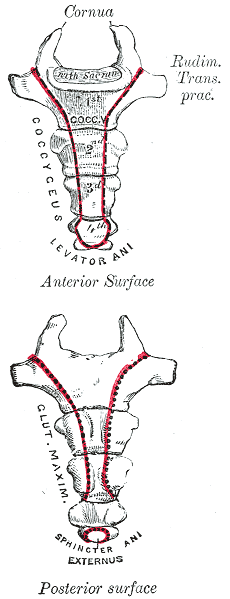
The coccyx ( : coccyges or coccyxes), commonly referred to as the tailbone, is the final segment of the vertebral column in all apes, and analogous structures in certain other mammals such as horses. In tailless primates (e.g. humans and other great apes) since '' Nacholapithecus'' (a Miocene hominoid),Nakatsukasa 2004, ''Acquisition of bipedalism'' (SeFig. 5entitled ''First coccygeal/caudal vertebra in short-tailed or tailless primates.''.) the coccyx is the remnant of a vestigial tail. In animals with bony tails, it is known as ''tailhead'' or ''dock'', in bird anatomy as ''tailfan''. It comprises three to five separate or fused coccygeal vertebrae below the sacrum, attached to the sacrum by a fibrocartilaginous joint, the sacrococcygeal symphysis, which permits limited movement between the sacrum and the coccyx. Structure The coccyx is formed of three, four or five rudimentary vertebrae. It articulates superiorly with the sacrum. In each of the first three segm ... [...More Info...] [...Related Items...] OR: [Wikipedia] [Google] [Baidu] |
Sacrum
The sacrum (plural: ''sacra'' or ''sacrums''), in human anatomy, is a large, triangular bone at the base of the spine that forms by the fusing of the sacral vertebrae (S1S5) between ages 18 and 30. The sacrum situates at the upper, back part of the pelvic cavity, between the two wings of the pelvis. It forms joints with four other bones. The two projections at the sides of the sacrum are called the alae (wings), and articulate with the ilium at the L-shaped sacroiliac joints. The upper part of the sacrum connects with the last lumbar vertebra (L5), and its lower part with the coccyx (tailbone) via the sacral and coccygeal cornua. The sacrum has three different surfaces which are shaped to accommodate surrounding pelvic structures. Overall it is concave (curved upon itself). The base of the sacrum, the broadest and uppermost part, is tilted forward as the sacral promontory internally. The central part is curved outward toward the posterior, allowing greater room for the ... [...More Info...] [...Related Items...] OR: [Wikipedia] [Google] [Baidu] |
Sacral Vertebra
The sacrum (plural: ''sacra'' or ''sacrums''), in human anatomy, is a large, triangular bone at the base of the spine that forms by the fusing of the sacral vertebrae (S1S5) between ages 18 and 30. The sacrum situates at the upper, back part of the pelvic cavity, between the two wings of the pelvis. It forms joints with four other bones. The two projections at the sides of the sacrum are called the alae (wings), and articulate with the ilium at the L-shaped sacroiliac joints. The upper part of the sacrum connects with the last lumbar vertebra (L5), and its lower part with the coccyx (tailbone) via the sacral and coccygeal cornua. The sacrum has three different surfaces which are shaped to accommodate surrounding pelvic structures. Overall it is concave (curved upon itself). The base of the sacrum, the broadest and uppermost part, is tilted forward as the sacral promontory internally. The central part is curved outward toward the posterior, allowing greater room for the pelv ... [...More Info...] [...Related Items...] OR: [Wikipedia] [Google] [Baidu] |
Sacrococcygeal Symphysis
The sacrococcygeal symphysis (sacrococcygeal articulation, articulation of the sacrum and coccyx) is an amphiarthrodial joint, formed between the oval surface at the apex of the sacrum, and the base of the coccyx. It is a slightly moveable jointMorris (2005), p 59 which is frequently, partially or completely, obliterated in old age,Palastanga (2006), p 334 homologous with the joints between the bodies of the vertebrae. Disc The sacrococcygeal disc or interosseus ligamentHuijbregts (2001), p 13 is similar to the intervertebral discs but thinner, thicker in front and behind than at the sides, and with a firmer texture. The articular surfaces are elliptical with longer transversal axes. The surface on the sacrum is convex and that on the coccyx concave. Occasionally the coccyx is freely movable on the sacrum, most notably during pregnancy; in such cases a synovial membrane is present. Ligaments The joint is strengthened by a series of ligaments: * The ventral or anterior s ... [...More Info...] [...Related Items...] OR: [Wikipedia] [Google] [Baidu] |
Levator Ani
The levator ani is a broad, thin muscle group, situated on either side of the pelvis. It is formed from three muscle components: the pubococcygeus, the iliococcygeus, and the puborectalis. It is attached to the inner surface of each side of the lesser pelvis, and these unite to form the greater part of the pelvic floor. The coccygeus muscle completes the pelvic floor, which is also called the ''pelvic diaphragm''. It supports the viscera in the pelvic cavity, and surrounds the various structures that pass through it. The levator ani is the main pelvic floor muscle and painfully contracts during vaginismus. It also contracts rhythmically during orgasm. Structure The levator ani is made up of 3 parts: * Iliococcygeus muscle * Pubococcygeus muscle * Puborectalis muscle The iliococcygeus arises from the inner side of the ischium (the lower and back part of the hip bone) and from the posterior part of the tendinous arch of the obturator fascia, and is attached to the coccyx and ... [...More Info...] [...Related Items...] OR: [Wikipedia] [Google] [Baidu] |
Vertebral Column
The vertebral column, also known as the backbone or spine, is part of the axial skeleton. The vertebral column is the defining characteristic of a vertebrate in which the notochord (a flexible rod of uniform composition) found in all chordates has been replaced by a segmented series of bone: vertebrae separated by intervertebral discs. Individual vertebrae are named according to their region and position, and can be used as anatomical landmarks in order to guide procedures such as lumbar punctures. The vertebral column houses the spinal canal, a cavity that encloses and protects the spinal cord. There are about 50,000 species of animals that have a vertebral column. The human vertebral column is one of the most-studied examples. Many different diseases in humans can affect the spine, with spina bifida and scoliosis being recognisable examples. The general structure of human vertebrae is fairly typical of that found in mammals, reptiles, and birds. The shape of the verte ... [...More Info...] [...Related Items...] OR: [Wikipedia] [Google] [Baidu] |
Vertebra
The spinal column, a defining synapomorphy shared by nearly all vertebrates, Hagfish are believed to have secondarily lost their spinal column is a moderately flexible series of vertebrae (singular vertebra), each constituting a characteristic irregular bone whose complex structure is composed primarily of bone, and secondarily of hyaline cartilage. They show variation in the proportion contributed by these two tissue types; such variations correlate on one hand with the cerebral/caudal rank (i.e., location within the backbone), and on the other with phylogenetic differences among the vertebrate taxa. The basic configuration of a vertebra varies, but the bone is its ''body'', with the central part of the body constituting the ''centrum''. The upper (closer to) and lower (further from), respectively, the cranium and its central nervous system surfaces of the vertebra body support attachment to the intervertebral discs. The posterior part of a vertebra forms a vertebral ... [...More Info...] [...Related Items...] OR: [Wikipedia] [Google] [Baidu] |
Spinous Process
The spinal column, a defining synapomorphy shared by nearly all vertebrates, Hagfish are believed to have secondarily lost their spinal column is a moderately flexible series of vertebrae (singular vertebra), each constituting a characteristic irregular bone whose complex structure is composed primarily of bone, and secondarily of hyaline cartilage. They show variation in the proportion contributed by these two tissue types; such variations correlate on one hand with the cerebral/caudal rank (i.e., location within the backbone), and on the other with phylogenetic differences among the vertebrate taxa. The basic configuration of a vertebra varies, but the bone is its ''body'', with the central part of the body constituting the ''centrum''. The upper (closer to) and lower (further from), respectively, the cranium and its central nervous system surfaces of the vertebra body support attachment to the intervertebral discs. The posterior part of a vertebra forms a vertebral a ... [...More Info...] [...Related Items...] OR: [Wikipedia] [Google] [Baidu] |
Lamina Of The Vertebral Arch
The spinal column, a defining synapomorphy shared by nearly all vertebrates,Hagfish are believed to have secondarily lost their spinal column is a moderately flexible series of vertebrae (singular vertebra), each constituting a characteristic irregular bone whose complex structure is composed primarily of bone, and secondarily of hyaline cartilage. They show variation in the proportion contributed by these two tissue types; such variations correlate on one hand with the cerebral/caudal rank (i.e., location within the backbone), and on the other with phylogenetic differences among the vertebrate taxa. The basic configuration of a vertebra varies, but the bone is its ''body'', with the central part of the body constituting the ''centrum''. The upper (closer to) and lower (further from), respectively, the cranium and its central nervous system surfaces of the vertebra body support attachment to the intervertebral discs. The posterior part of a vertebra forms a vertebral arch ( ... [...More Info...] [...Related Items...] OR: [Wikipedia] [Google] [Baidu] |
Transverse Process
The spinal column, a defining synapomorphy shared by nearly all vertebrates, Hagfish are believed to have secondarily lost their spinal column is a moderately flexible series of vertebrae (singular vertebra), each constituting a characteristic irregular bone whose complex structure is composed primarily of bone, and secondarily of hyaline cartilage. They show variation in the proportion contributed by these two tissue types; such variations correlate on one hand with the cerebral/caudal rank (i.e., location within the backbone), and on the other with phylogenetic differences among the vertebrate taxa. The basic configuration of a vertebra varies, but the bone is its ''body'', with the central part of the body constituting the ''centrum''. The upper (closer to) and lower (further from), respectively, the cranium and its central nervous system surfaces of the vertebra body support attachment to the intervertebral discs. The posterior part of a vertebra forms a vertebral a ... [...More Info...] [...Related Items...] OR: [Wikipedia] [Google] [Baidu] |
Spinal Cord
The spinal cord is a long, thin, tubular structure made up of nervous tissue, which extends from the medulla oblongata in the brainstem to the lumbar region of the vertebral column (backbone). The backbone encloses the central canal of the spinal cord, which contains cerebrospinal fluid. The brain and spinal cord together make up the central nervous system (CNS). In humans, the spinal cord begins at the occipital bone, passing through the foramen magnum and then enters the spinal canal at the beginning of the cervical vertebrae. The spinal cord extends down to between the first and second lumbar vertebrae, where it ends. The enclosing bony vertebral column protects the relatively shorter spinal cord. It is around long in adult men and around long in adult women. The diameter of the spinal cord ranges from in the cervical and lumbar regions to in the thoracic area. The spinal cord functions primarily in the transmission of nerve signals from the motor cortex to the b ... [...More Info...] [...Related Items...] OR: [Wikipedia] [Google] [Baidu] |
Anterior Sacrococcygeal Ligament
The anterior sacrococcygeal ligament or ventral sacrococcygeal ligament consists of a few irregular fibers, which descend from the anterior surface of the sacrum to the front of the coccyx, blending with the periosteum.Morris (2005), p 59 This shortSinnatamby (2006), p 336 ligament forms the continuation of the anterior longitudinal ligament and stretches over the sacrococcygeal symphysis.OMD: DefinitionJinkins (2000), p 538Ebrall (2004), 243 See also * Posterior sacrococcygeal ligament * Coccydynia (coccyx pain, tailbone pain) * Ganglion impar The pelvic portion of each sympathetic trunk is situated in front of the sacrum The sacrum (plural: ''sacra'' or ''sacrums''), in human anatomy, is a large, triangular bone at the base of the spine that forms by the fusing of the sacral ver ... Notes References * * * * Ligaments of the torso Bones of the vertebral column {{ligament-stub ... [...More Info...] [...Related Items...] OR: [Wikipedia] [Google] [Baidu] |


