|
Septum
In biology, a septum (Latin for ''something that encloses''; plural septa) is a wall, dividing a cavity or structure into smaller ones. A cavity or structure divided in this way may be referred to as septate. Examples Human anatomy * Interatrial septum, the wall of tissue that is a sectional part of the left and right atria of the heart * Interventricular septum, the wall separating the left and right ventricles of the heart * Lingual septum, a vertical layer of fibrous tissue that separates the halves of the tongue. * Nasal septum: the cartilage wall separating the nostrils of the nose * Alveolar septum: the thin wall which separates the alveoli from each other in the lungs * Orbital septum, a palpebral ligament in the upper and lower eyelids * Septum pellucidum or septum lucidum, a thin structure separating two fluid pockets in the brain * Uterine septum, a malformation of the uterus * Vaginal septum, a lateral or transverse partition inside the vagina * Intermuscu ... [...More Info...] [...Related Items...] OR: [Wikipedia] [Google] [Baidu] |
Interventricular Septum
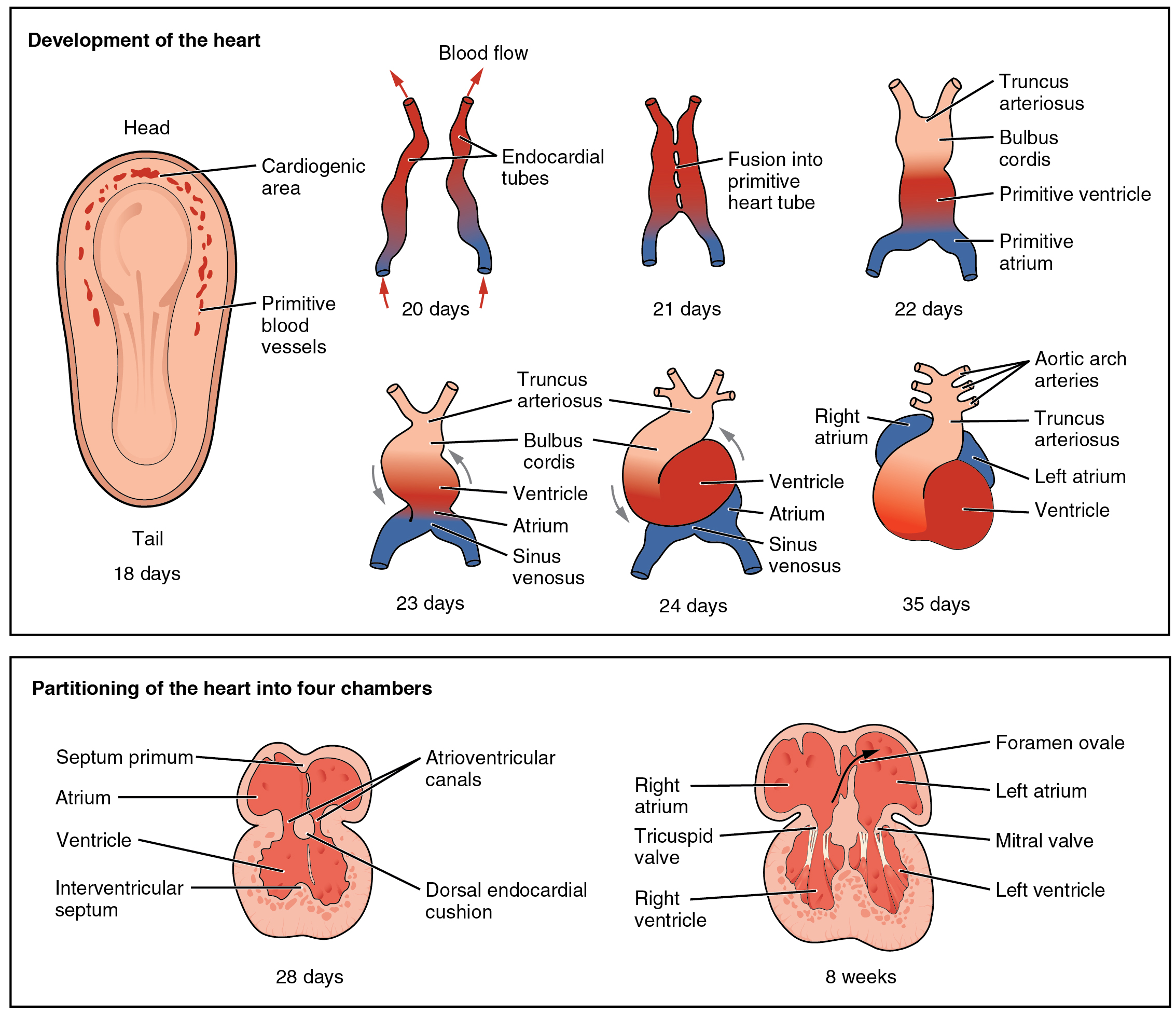
The interventricular septum (IVS, or ventricular septum, or during development septum inferius) is the stout wall separating the ventricles, the lower chambers of the heart, from one another. The ventricular septum is directed obliquely backward to the right and curved with the convexity toward the right ventricle; its margins correspond with the anterior and posterior interventricular sulci. The lower part of the septum, which is the major part, is thick and muscular, and its much smaller upper part is thin and membraneous. During each cardiac cycle the interventricular septum contracts by shortening longitudinally and becoming thicker. Structure The interventricular septum is the stout wall separating the ventricles, the lower chambers of the heart, from one another. The ventricular septum is directed obliquely backward to the right and curved with the convexity toward the right ventricle; its margins correspond with the anterior and posterior longitudinal sulci. Th ... [...More Info...] [...Related Items...] OR: [Wikipedia] [Google] [Baidu] |
Interatrial Septum
The interatrial septum is the wall of tissue that separates the right and left atria of the heart. Structure The interatrial septum is a that lies between the left atrium and right atrium of the human heart. The interatrial septum lies at angle of 65 degrees from right posterior to left anterior because right atrium is located at the right side of the body while left atrium is located at the left side of the body. Development The interatrial septum forms during the first and second months of fetal development. Formation of the septum occurs in several stages. The first is the development of the septum primum, a crescent-shaped piece of tissue forming the initial divider between the right and left atria. Because of its crescent shape, the septum primum does not fully occlude the space between the left and right atria; the opening that remains is called the ostium primum. During fetal development, this opening allows blood to be shunted from the right atrium to the left. As th ... [...More Info...] [...Related Items...] OR: [Wikipedia] [Google] [Baidu] |
Nasal Septum
The nasal septum () separates the left and right airways of the nasal cavity, dividing the two nostrils. It is depressed by the depressor septi nasi muscle. Structure The fleshy external end of the nasal septum is called the columella or columella nasi, and is made up of cartilage and soft tissue. The nasal septum contains bone and hyaline cartilage. It is normally about 2 mm thick. The nasal septum is composed of four structures: * Perpendicular plate of ethmoid bone * Vomer bone * Septal nasal cartilage * Maxillary bone (the crest) The lowest part of the septum is a narrow strip of bone that projects from the maxilla and the palatine bones, and is the length of the septum. This strip of bone is called the maxillary crest; it articulates in front with the septal nasal cartilage, and at the back with the vomer. The maxillary crest is described in the anatomy of the nasal septum as having a maxillary component and a palatine component. Development At an early perio ... [...More Info...] [...Related Items...] OR: [Wikipedia] [Google] [Baidu] |
Uterine Septum
A uterine septum is a form of a congenital malformation where the uterine cavity is partitioned by a longitudinal septum; the outside of the uterus has a normal typical shape. The wedge-like partition may involve only the superior part of the cavity resulting in an ''incomplete septum'' or a ''subseptate uterus'', or less frequently the total length of the cavity (''complete septum'') and the cervix resulting in a double cervix. The septation may also continue caudally into the vagina resulting in a " double vagina". Signs and symptoms The condition may not be known to the affected individual and not result in any reproductive problems; thus normal pregnancies may occur. In more serious cases have reported high infertility rates. However, it is associated with a higher risk for miscarriage, premature birth Preterm birth, also known as premature birth, is the birth of a baby at fewer than 37 weeks gestational age, as opposed to full-term delivery at approximately 40 weeks. E ... [...More Info...] [...Related Items...] OR: [Wikipedia] [Google] [Baidu] |
Orbital Septum
In anatomy, the orbital septum (palpebral fascia) is a membranous sheet that acts as the anterior (frontal) boundary of the orbit. It extends from the orbital rims to the eyelids. It forms the fibrous portion of the eyelids. Structure In the upper eyelid, the orbital septum blends with the tendon of the levator palpebrae superioris, and in the lower eyelid with the tarsal plate. When the eyes are closed, the whole orbital opening is covered by the septum and tarsi. Medially it is thin, and, becoming separated from the medial palpebral ligament, attaches to the lacrimal bone at its posterior crest. The medial ligament and its much weaker lateral counterpart, attached to the septum and orbit, keep the lids stable as the eye moves. The septum is perforated by the vessels and nerves which pass from the orbital cavity to the face and scalp. Clinical significance Orbital septum supports the orbital contents located posterior to it, especially orbital fat. The septum can be wea ... [...More Info...] [...Related Items...] OR: [Wikipedia] [Google] [Baidu] |
Septate Hyphae Of The Fungus Alternaria Sp
In biology, a septum (Latin for ''something that encloses''; plural septa) is a wall, dividing a cavity or structure into smaller ones. A cavity or structure divided in this way may be referred to as septate. Examples Human anatomy * Interatrial septum, the wall of tissue that is a sectional part of the left and right atria of the heart * Interventricular septum, the wall separating the left and right ventricles of the heart * Lingual septum, a vertical layer of fibrous tissue that separates the halves of the tongue. * Nasal septum: the cartilage wall separating the nostrils of the nose * Alveolar septum: the thin wall which separates the alveoli from each other in the lungs * Orbital septum, a palpebral ligament in the upper and lower eyelids * Septum pellucidum or septum lucidum, a thin structure separating two fluid pockets in the brain * Uterine septum, a malformation of the uterus * Vaginal septum, a lateral or transverse partition inside the vagina * Intermuscu ... [...More Info...] [...Related Items...] OR: [Wikipedia] [Google] [Baidu] |
Ventricular Septal Defect
A ventricular septal defect (VSD) is a defect in the ventricular septum, the wall dividing the left and right ventricles of the heart. The extent of the opening may vary from pin size to complete absence of the ventricular septum, creating one common ventricle. The ventricular septum consists of an inferior muscular and superior membranous portion and is extensively innervated with conducting cardiomyocytes. The membranous portion, which is close to the atrioventricular node, is most commonly affected in adults and older children in the United States. It is also the type that will most commonly require surgical intervention, comprising over 80% of cases. Membranous ventricular septal defects are more common than muscular ventricular septal defects, and are the most common congenital cardiac anomaly. Signs and symptoms Ventricular septal defect is usually symptomless at birth. It usually manifests a few weeks after birth. VSD is an acyanotic congenital heart defect, aka a l ... [...More Info...] [...Related Items...] OR: [Wikipedia] [Google] [Baidu] |
Septum Pellucidum
The septum pellucidum (Latin for "translucent wall") is a thin, triangular, vertical double membrane separating the anterior horns of the left and right lateral ventricles of the brain. It runs as a sheet from the corpus callosum down to the fornix. The septum is not present in the syndrome septo-optic dysplasia. Structure The septum pellucidum is located in the septal area in the midline of the brain between the two cerebral hemispheres. The septal area is also the location of the septal nuclei. It is attached to the lower part of the corpus callosum, the large collection of nerve fibers that connect the two cerebral hemispheres. It is attached to the front forward part of the fornix. The lateral ventricles sit on either side of the septum. The septum pellucidum consists of two layers or ''laminae'' of both white and gray matter. During fetal development, there is a space between the two laminae called the cave of septum pellucidum that, in ninety percent of cases, disapp ... [...More Info...] [...Related Items...] OR: [Wikipedia] [Google] [Baidu] |
Vaginal Septum
A vaginal septum is a vaginal anomaly that is partition within the vagina; such a septum could be either longitudinal or transverse. In some affected women, the septum is partial or does not extend the length or width of the vagina. Pain during intercourse can be a symptom. A longitudinal vaginal septum develops during embryogenesis when there is an incomplete fusion of the lower parts of the two Müllerian ducts. As a result, there may appear to be two openings to the vagina. There may be associated duplications of the more cranial parts of the Müllerian derivatives, a double cervix, and either a uterine septum or uterus didelphys (double uterus). A transverse septum forms during embryogenesis when the Müllerian ducts do not fuse to the urogenital sinus. A complete transverse septum can occur across the vagina at different levels. Menstrual flow can be blocked, and is a cause of primary amenorrhea. The accumulation of menstrual debris behind the septum is termed cryptome ... [...More Info...] [...Related Items...] OR: [Wikipedia] [Google] [Baidu] |
Septum (cell Biology)
A septum in cell biology is the new cell wall that forms between two daughter cells as a result of cell division. In yeast, septins form a ring structure, to which other proteins are recruited. In particular, chitinase 2 is required, an enzyme that synthesises chitin Chitin ( C8 H13 O5 N)n ( ) is a long-chain polymer of ''N''-acetylglucosamine, an amide derivative of glucose. Chitin is probably the second most abundant polysaccharide in nature (behind only cellulose); an estimated 1 billion tons of chit ... thereby building up the primary septum. A secondary septum of β-glucans and mannoproteins is then assembled using the enzyme 1,3-Beta-glucan synthase, and the primary septum degraded during cell separation. After degradation of the primary septum, a chitinous bud scar remains on both the mother and daughter cell. Composition In Schizosaccharomyces pombe, the primary septum is composed of linear β(1,3)-D-glucan, β(1,6) branches, and α(1,3)-D-glucan. The sec ... [...More Info...] [...Related Items...] OR: [Wikipedia] [Google] [Baidu] |
Intermuscular Septum (other)
Intermuscular septum can refer to: * medial intermuscular septum of arm * medial intermuscular septum of thigh * lateral intermuscular septum of arm * lateral intermuscular septum of thigh * anterior intermuscular septum of leg * posterior intermuscular septum of leg {{disambig ... [...More Info...] [...Related Items...] OR: [Wikipedia] [Google] [Baidu] |
Lingual Septum
The lingual septum consists of a vertical layer of fibrous tissue, extending throughout the entire length of the median plane of the tongue, though not quite reaching the dorsum. The lingual septum is closely associated with the hyoglossus membrane, allowing the binding of the tongue to the hyoid muscles. Visualization through implementing a vertical groove along the tongue called the median sulcus. It is thicker behind than in front, and occasionally contains a small fibrocartilage, about 6 mm. in length. See also *Tongue piercing *Tongue splitting *Mouth cavity *Tongue *Fibrous tissue Fiber or fibre (from la, fibra, links=no) is a natural or artificial substance that is significantly longer than it is wide. Fibers are often used in the manufacture of other materials. The strongest engineering materials often incorporate ... References . Tongue {{anatomy-stub ... [...More Info...] [...Related Items...] OR: [Wikipedia] [Google] [Baidu] |


