|
Intestinal Mucosa
The gastrointestinal wall of the gastrointestinal tract is made up of four layers of specialised tissue. From the inner cavity of the gut (the lumen) outwards, these are: # Mucosa # Submucosa # Muscular layer # Serosa or adventitia The mucosa is the innermost layer of the gastrointestinal tract. It surrounds the lumen of the tract and comes into direct contact with digested food (chyme). The mucosa itself is made up of three layers: the epithelium, where most digestive, absorptive and secretory processes occur; the lamina propria, a layer of connective tissue, and the muscularis mucosae, a thin layer of smooth muscle. The submucosa contains nerves including the submucous plexus (also called Meissner's plexus), blood vessels and elastic fibres with collagen, that stretches with increased capacity but maintains the shape of the intestine. The muscular layer surrounds the submucosa. It comprises layers of smooth muscle in longitudinal and circular orientation that also helps w ... [...More Info...] [...Related Items...] OR: [Wikipedia] [Google] [Baidu] |
Human Gastrointestinal Tract
The gastrointestinal tract (GI tract, digestive tract, alimentary canal) is the tract or passageway of the digestive system that leads from the mouth to the anus. The GI tract contains all the major organs of the digestive system, in humans and other animals, including the esophagus, stomach, and intestines. Food taken in through the mouth is digested to extract nutrients and absorb energy, and the waste expelled at the anus as feces. ''Gastrointestinal'' is an adjective meaning of or pertaining to the stomach and intestines. Most animals have a "through-gut" or complete digestive tract. Exceptions are more primitive ones: sponges have small pores ( ostia) throughout their body for digestion and a larger dorsal pore ( osculum) for excretion, comb jellies have both a ventral mouth and dorsal anal pores, while cnidarians and acoels have a single pore for both digestion and excretion. The human gastrointestinal tract consists of the esophagus, stomach, and intestines, and is ... [...More Info...] [...Related Items...] OR: [Wikipedia] [Google] [Baidu] |
Mucus
Mucus ( ) is a slippery aqueous secretion produced by, and covering, mucous membranes. It is typically produced from cells found in mucous glands, although it may also originate from mixed glands, which contain both serous and mucous cells. It is a viscous colloid containing inorganic salts, antimicrobial enzymes (such as lysozymes), immunoglobulins (especially IgA), and glycoproteins such as lactoferrin and mucins, which are produced by goblet cells in the mucous membranes and submucosal glands. Mucus serves to protect epithelial cells in the linings of the respiratory, digestive, and urogenital systems, and structures in the visual and auditory systems from pathogenic fungi, bacteria and viruses. Most of the mucus in the body is produced in the gastrointestinal tract. Amphibians, fish, snails, slugs, and some other invertebrates also produce external mucus from their epidermis as protection against pathogens, and to help in movement and is also produced in fish ... [...More Info...] [...Related Items...] OR: [Wikipedia] [Google] [Baidu] |
Catastalsis
Catastalsis is the rhythmic contraction and relaxation of the muscle of the intestines. It resembles ordinary peristalsis Peristalsis ( , ) is a radially symmetrical contraction and relaxation of muscles that propagate in a wave down a tube, in an anterograde direction. Peristalsis is progression of coordinated contraction of involuntary circular muscles, whi ... but is not preceded by a wave of inhibition. References Digestive system {{digestive-stub ... [...More Info...] [...Related Items...] OR: [Wikipedia] [Google] [Baidu] |
Muscularis Mucosa
The lamina muscularis mucosae (or muscularis mucosae) is a thin layer ( lamina) of muscle of the gastrointestinal tract, located outside the lamina propria, and separating it from the submucosa. It is present in a continuous fashion from the esophagus to the upper rectum (the exact nomenclature of the rectum's muscle layers is still being debated). A discontinuous muscularis mucosae–like muscle layer is present in the urinary tract, from the renal pelvis to the bladder; as it is discontinuous, it should not be regarded as a true muscularis mucosae. In the gastrointestinal tract, the term ''mucosa'' or ''mucous membrane'' refers to the combination of epithelium, lamina propria, and (where it occurs) muscularis mucosae.H.G. Burkitt et al., ''Wheater's Functional Histology, 3rd ed.'' The etymology suggests this, since the Latin names translate to "the mucosa's own special layer" (''lamina propria mucosae'') and "muscular layer of the mucosa" (''lamina muscularis mucosae''). ... [...More Info...] [...Related Items...] OR: [Wikipedia] [Google] [Baidu] |
Intestinal Gland
In histology, an intestinal gland (also crypt of Lieberkühn and intestinal crypt) is a gland found in between villi in the intestinal epithelium lining of the small intestine and large intestine (or colon). The glands and intestinal villi are covered by epithelium, which contains multiple types of cells: enterocytes (absorbing water and electrolytes), goblet cells (secreting mucus), enteroendocrine cells (secreting hormones), cup cells, tuft cells, and at the base of the gland, Paneth cells (secreting anti-microbial peptides) and stem cells. Structure Intestinal glands are found in the epithelia of the small intestine, namely the duodenum, jejunum, and ileum, and in the large intestine (colon), where they are sometimes called ''colonic crypts''. Intestinal glands of the small intestine contain a base of replicating stem cells, Paneth cells of the innate immune system, and goblet cells, which produce mucus. In the colon, crypts do not have Paneth cells. Function The enterocyte ... [...More Info...] [...Related Items...] OR: [Wikipedia] [Google] [Baidu] |
Lipid
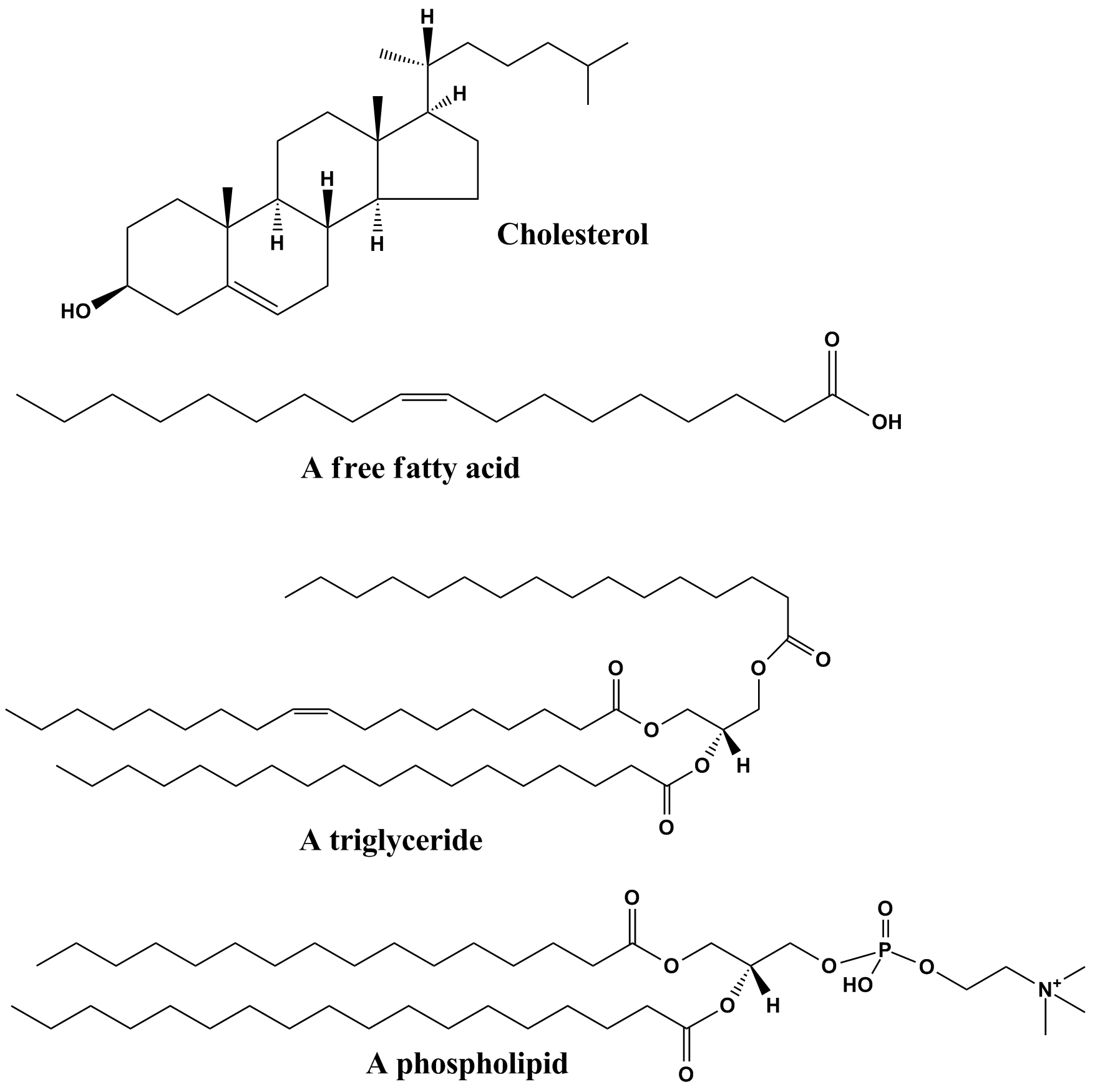
Lipids are a broad group of naturally-occurring molecules which includes fats, waxes, sterols, fat-soluble vitamins (such as vitamins A, D, E and K), monoglycerides, diglycerides, phospholipids, and others. The functions of lipids include storing energy, signaling, and acting as structural components of cell membranes. Lipids have applications in the cosmetic and food industries, and in nanotechnology. Lipids may be broadly defined as hydrophobic or amphiphilic small molecules; the amphiphilic nature of some lipids allows them to form structures such as vesicles, multilamellar/unilamellar liposomes, or membranes in an aqueous environment. Biological lipids originate entirely or in part from two distinct types of biochemical subunits or "building-blocks": ketoacyl and isoprene groups. Using this approach, lipids may be divided into eight categories: fatty acyls, glycerolipids, glycerophospholipids, sphingolipids, saccharolipids, and polyketides (derived from cond ... [...More Info...] [...Related Items...] OR: [Wikipedia] [Google] [Baidu] |
Lymphatic System
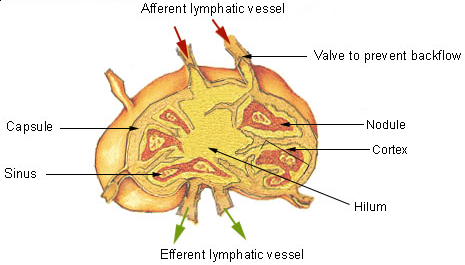
The lymphatic system, or lymphoid system, is an organ system in vertebrates that is part of the immune system, and complementary to the circulatory system. It consists of a large network of lymphatic vessels, lymph nodes, lymphatic or lymphoid organs, and lymphoid tissues. The vessels carry a clear fluid called lymph (the Latin word ''lympha'' refers to the deity of fresh water, "Lympha") back towards the heart, for re-circulation. Unlike the circulatory system that is a closed system, the lymphatic system is open. The human circulatory system processes an average of 20 litres of blood per day through capillary filtration, which removes plasma from the blood. Roughly 17 litres of the filtered blood is reabsorbed directly into the blood vessels, while the remaining three litres are left in the interstitial fluid. One of the main functions of the lymphatic system is to provide an accessory return route to the blood for the surplus three litres. The other main function is that ... [...More Info...] [...Related Items...] OR: [Wikipedia] [Google] [Baidu] |
Lacteal
A lacteal is a lymphatic capillary that absorbs dietary fats in the villi of the small intestine. Triglycerides are emulsified by bile and hydrolyzed by the enzyme lipase, resulting in a mixture of fatty acids, di- and monoglycerides. These then pass from the intestinal lumen into the enterocyte, where they are re-esterified to form triglyceride. The triglyceride is then combined with phospholipids, cholesterol ester, and apolipoprotein B48 to form chylomicrons. These chylomicrons then pass into the lacteals, forming a milky substance known as chyle. The lacteals merge to form larger lymphatic vessels that transport the chyle to the thoracic duct where it is emptied into the bloodstream at the subclavian vein. At this point, the fats are in the bloodstream in the form of chylomicrons. Once in the blood, chylomicrons are subject to delipidation by lipoprotein lipase. Eventually, enough lipid has been lost and additional apolipoproteins gained, that the resulting particle ... [...More Info...] [...Related Items...] OR: [Wikipedia] [Google] [Baidu] |
Intestinal Villus
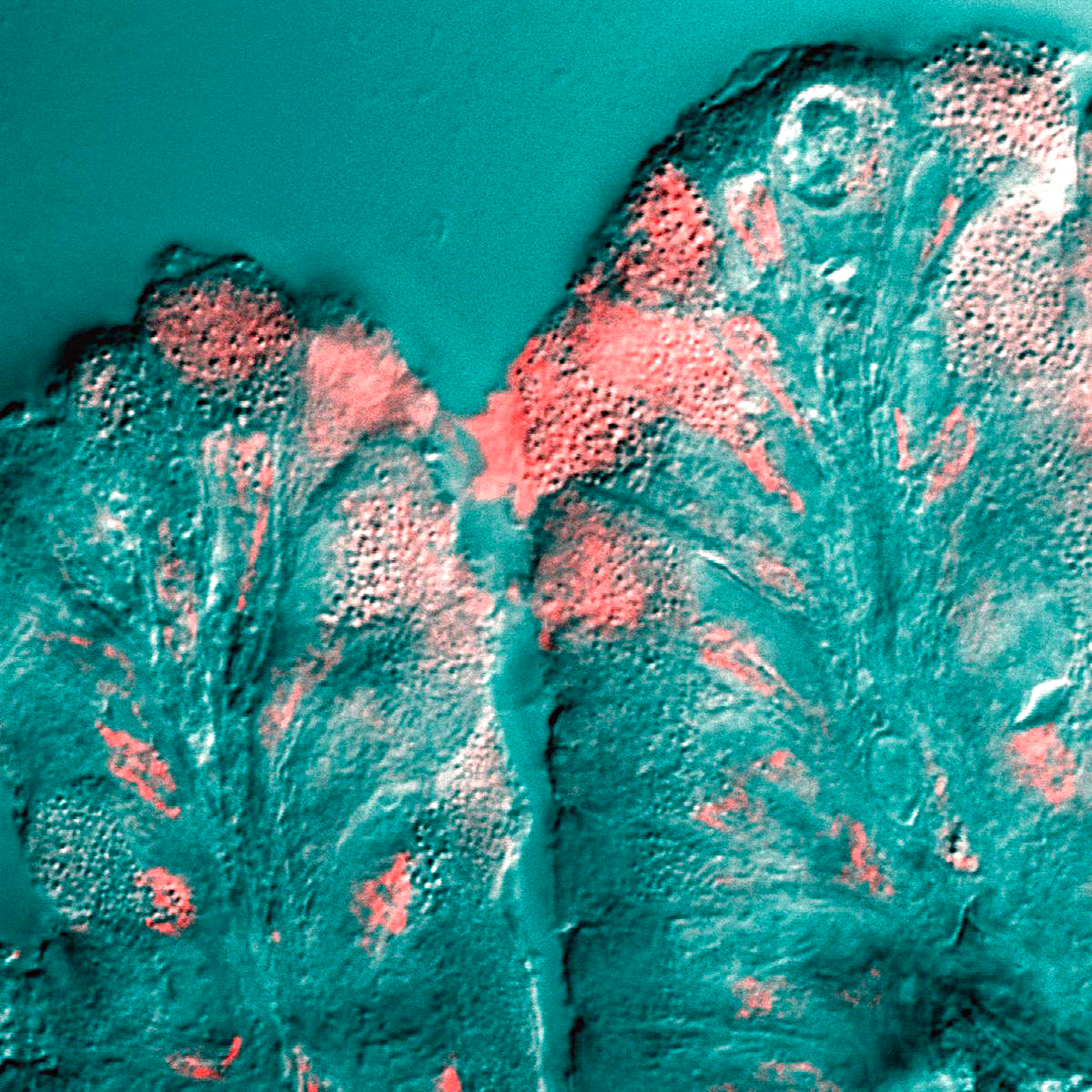
Intestinal villi (singular: villus) are small, finger-like projections that extend into the lumen of the small intestine. Each villus is approximately 0.5–1.6 mm in length (in humans), and has many microvilli projecting from the enterocytes of its epithelium which collectively form the striated or brush border. Each of these microvilli are about 1 µm in length, around 1000 times shorter than a single villus. The intestinal villi are much smaller than any of the circular folds in the intestine. Villi increase the internal surface area of the intestinal walls making available a greater surface area for absorption. An increased absorptive area is useful because digested nutrients (including monosaccharide and amino acids) pass into the semipermeable villi through diffusion, which is effective only at short distances. In other words, increased surface area (in contact with the fluid in the lumen) decreases the average distance travelled by nutrient molecules, so effect ... [...More Info...] [...Related Items...] OR: [Wikipedia] [Google] [Baidu] |
Goblet Cell
Goblet cells are simple columnar epithelial cells that secrete gel-forming mucins, like mucin 5AC. The goblet cells mainly use the merocrine method of secretion, secreting vesicles into a duct, but may use apocrine methods, budding off their secretions, when under stress. The term '' goblet'' refers to the cell's goblet-like shape. The apical portion is shaped like a cup, as it is distended by abundant mucus laden granules; its basal portion lacks these granules and is shaped like a stem. The goblet cell is highly polarized with the nucleus and other organelles concentrated at the base of the cell and secretory granules containing mucin, at the apical surface. The apical plasma membrane projects short microvilli to give an increased surface area for secretion. Goblet cells are typically found in the respiratory, reproductive and gastrointestinal tracts and are surrounded by other columnar cells. Biased differentiation of airway basal cells in the respiratory epithelium, in ... [...More Info...] [...Related Items...] OR: [Wikipedia] [Google] [Baidu] |
Glandular Epithelium
Epithelium or epithelial tissue is one of the four basic types of animal tissue, along with connective tissue, muscle tissue and nervous tissue. It is a thin, continuous, protective layer of compactly packed cells with a little intercellular matrix. Epithelial tissues line the outer surfaces of organs and blood vessels throughout the body, as well as the inner surfaces of cavities in many internal organs. An example is the epidermis, the outermost layer of the skin. There are three principal shapes of epithelial cell: squamous (scaly), columnar, and cuboidal. These can be arranged in a singular layer of cells as simple epithelium, either squamous, columnar, or cuboidal, or in layers of two or more cells deep as stratified (layered), or ''compound'', either squamous, columnar or cuboidal. In some tissues, a layer of columnar cells may appear to be stratified due to the placement of the nuclei. This sort of tissue is called pseudostratified. All glands are made up of epithel ... [...More Info...] [...Related Items...] OR: [Wikipedia] [Google] [Baidu] |


_and_foveolar_cells_in_incomplete_Barrett's_esophagus.jpg)
